Welcome to China Oncology,
China Oncology ›› 2025, Vol. 35 ›› Issue (5): 449-456.doi: 10.19401/j.cnki.1007-3639.2025.05.003
• Original article • Previous Articles Next Articles
YING Leilei( ), LI Kening, CHEN Chao, WANG Ying, HUANG Haozhe, WANG Biao, LI Wentao, HE Xinhong()
Received:2025-03-25
Revised:2025-05-09
Online:2025-05-30
Published:2025-06-10
Contact:
HE Xinhong
Supported by:Share article
CLC Number:
YING Leilei, LI Kening, CHEN Chao, WANG Ying, HUANG Haozhe, WANG Biao, LI Wentao, HE Xinhong. Impact of tumor diameter on post-radiofrequency ablation survival and local progression risk in patients with colorectal cancer lung metastasis[J]. China Oncology, 2025, 35(5): 449-456.
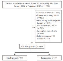
Fig. 1
Flowchart of patient inclusion and exclusion"
Tab. 1
Clinical characteristics of patients enrolled [n (%)]"
| Characteristic | Total (n=134) | Small group (n=77) | Large group (n=57) | P value |
|---|---|---|---|---|
| Age/year $\bar{x} \pm s$ | 59.3±12.1 | 57.6±11.7 | 60.3±12.3 | 0.226 |
| Gender | 0.714 | |||
| Female | 54 (40.3) | 30 (39.0) | 24 (42.1) | |
| Male | 80 (59.7) | 47 (61.0) | 33 (57.9) | |
| Location | 0.494 | |||
| Left | 73 (54.5) | 40 (51.9) | 33 (57.9) | |
| Right | 61 (45.5) | 37 (48.1) | 24 (42.1) | |
| Number of lesions | 0.833 | |||
| 1 | 107 (79.9) | 61 (79.2) | 46 (80.7) | |
| 2 | 27 (20.1) | 16 (20.8) | 11 (19.3) | |
| Extrapulmonary metastatic | 0.62 | |||
| None | 89 (66.4) | 53 (68.8) | 36 (63.2) | |
| Exist | 45 (33.6) | 24 (31.2) | 21 (36.8) | |
| Preoperative CEA/(ng·mL-1) | 0.601 | |||
| <5 | 69 (51.5) | 39 (50.6) | 30 (52.6) | |
| ≥5 | 65 (48.5) | 38 (49.4) | 27 (47.4) |

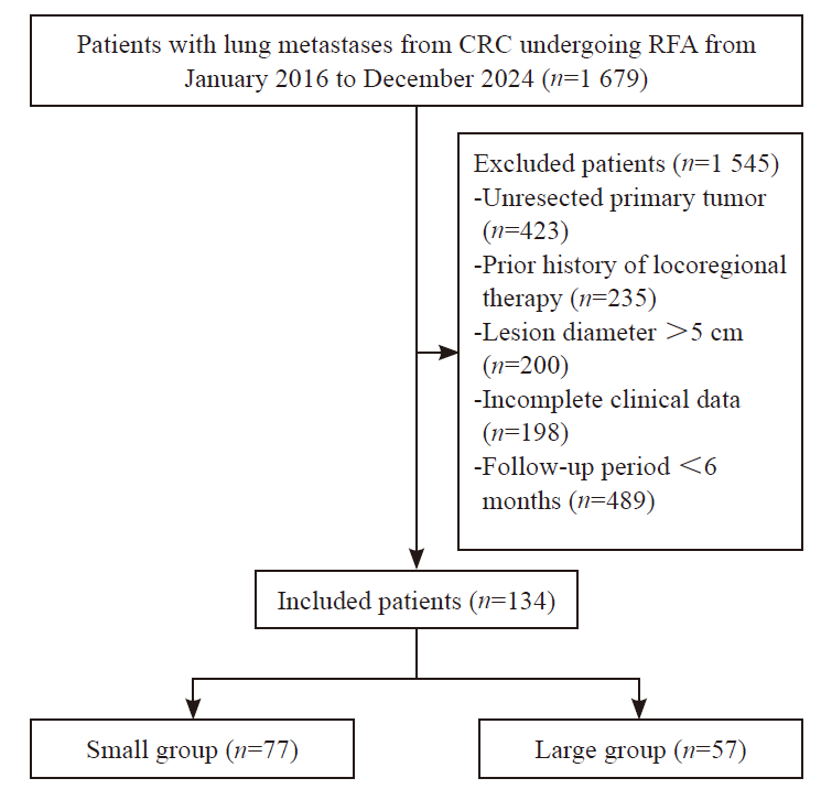
Fig. 2
OS rate curves of CRC patients with pulmonary metastases of different tumor diameters after RFA"
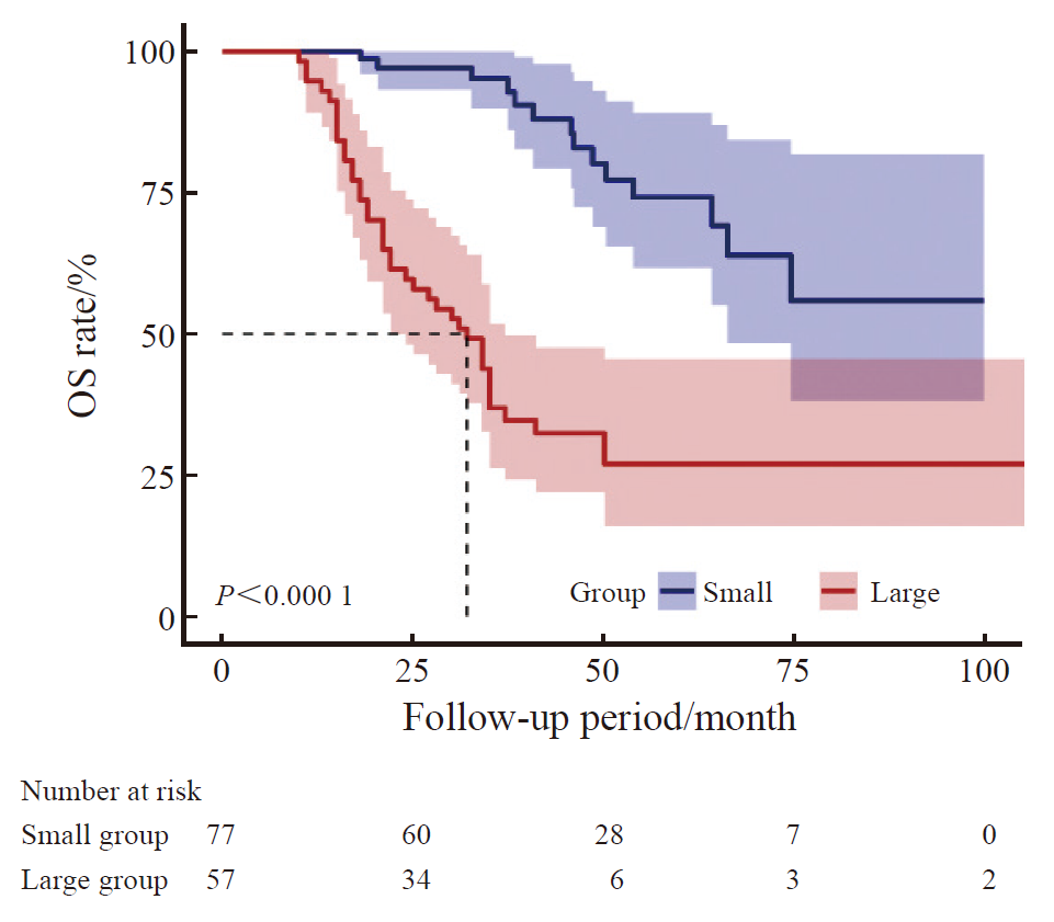
Fig. 3
PFS rate curves of CRC patients with pulmonary metastases of different tumor diameters after RFA"


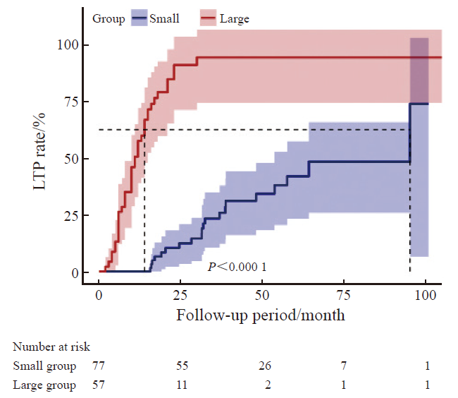
Fig. 4
LTP rate curves of CRC patients with pulmonary metastases of different tumor diameters after RFA"
Tab. 2
Univariate analysis of the correlation between RFA and OS in CRC patients with lung metastases"
| Variable | β (95% CI) | P value |
|---|---|---|
| Age | 1.01 (0.99, 1.04) | 0.280 |
| Gender | 0.366 | |
| Female | 1 (reference) | |
| Male | 0.78 (0.45, 1.34) | |
| Location | 0.849 | |
| Right | 1 (reference) | |
| Left | 0.95 (0.55, 1.63) | |
| Diameter | 1.96 (1.55, 2.48) | <0.001 |
| Preoperative CEA/(ng·mL-1) | 0.033 | |
| <5 | 1 (reference) | |
| ≥5 | 1.82 (1.05, 3.17) | |
| Extrapulmonary metastasis | 0.298 | |
| None | 1 (reference) | |
| Exist | 1.26 (0.81, 1.96) | |
| Number of lesions | 0.300 | |
| 1 | 1 (reference) | |
| 2 | 0.66 (0.29, 1.46) |
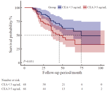
Fig. 5
Survival curves of CRC patients with lung metastasis after RFA treatment stratified by preoperative CEA levels"

Tab. 3
Multivariable COX regression analysis of the correlation between different tumor diameters and OS in patients with CRC pulmonary metastases following RFA"
| Group | Total | Event | Crude | Model-1 | Model-2 | Model-3 |
|---|---|---|---|---|---|---|
| Small | 77 | 14 | 1 (reference) | 1 (reference) | 1 (reference) | 1 (reference) |
| Large | 57 | 39 | 5.7 (3.06-10.6) | 5.84 (3.09-11.05) | 6.39 (3.34-12.21) | 6.49 (3.18-13.24) |
| P value | <0.001 | <0.001 | <0.001 | <0.001 |
| [1] | BRAY F, LAVERSANNE M, SUNG H, et al. Global cancer statistics 2022: GLOBOCAN estimates of incidence and mortality worldwide for 36 cancers in 185 countries[J]. CA Cancer J Clin, 2024, 74(3): 229-263. |
| [2] | WANG Z H, WANG X C, YUAN J J, et al. Survival benefit of palliative local treatments and efficacy of different pharmacotherapies in colorectal cancer with lung metastasis: results from a large retrospective study[J]. Clin Colorectal Cancer, 2018, 17(2): e233-e255. |
| [3] | 金晶, 顾晋, 沈琳. 结直肠癌肺转移多学科综合治疗专家共识(2018版)[J]. 实用肿瘤杂志, 2018, 33(6): 487-501. |
| JIN J, GU J, SHEN L. Expert consensus on multidisciplinary therapy of colorectal cancer with lung metastases (2018 edition)[J]. J Pract Oncol, 2018, 33(6): 487-501. | |
| [4] | 叶欣, 范卫君, 王徽, 等. 热消融治疗原发性和转移性肺部肿瘤专家共识(2017年版)[J]. 中国肺癌杂志, 2017, 20(7): 433-445. |
| YE X, FAN W J, WANG H, et al. Expert consensus for thermal ablation of primary and metastatic lung tumors (2017 edition)[J]. Chin J Lung Cancer, 2017, 20(7): 433-445. | |
| [5] | CERVANTES A, ADAM R, ROSELLÓ S, et al. Metastatic colorectal cancer: ESMO clinical practice guideline for diagnosis, treatment and follow-up[J]. Ann Oncol, 2023, 34(1): 10-32. |
| [6] | 中华医学会肿瘤学分会, 国家卫生健康委员会医政司. 中国结直肠癌诊疗规范(2023版)[J]. 协和医学杂志, 2023, 14(4): 706-733. |
| Oncology Branch of Chinese Medical Association; Medical Administration Bureau, National Health Commission. Chinese guidelines for the diagnosis and treatment of colorectal cancer (2023 edition)[J]. Med J Peking Union Med Coll Hosp, 2023, 14(04): 706-733. | |
| [7] | DE BAÈRE T, AUPÉRIN A, DESCHAMPS F, et al. Radiofrequency ablation is a valid treatment option for lung metastases: experience in 566 patients with 1 037 metastases[J]. Ann Oncol, 2015, 26(5): 987-991. |
| [8] | SHEN C, TANNENBAUM D, HORN R, et al. Overall survival in phase 3 clinical trials and the surveillance, epidemiology, and end results database in patients with metastatic colorectal cancer, 1986-2016: a systematic review[J]. JAMA Netw Open, 2022, 5(5): e2213588. |
| [9] | ENG C, YOSHINO T, RUÍZ-GARCÍA E, et al. Colorectal cancer[J]. Lancet, 2024, 404(10449): 294-310. |
| [10] | HUANG B Y, ZHOU J J, YU Z Q, et al. Long-term observation of CT-guided radiofrequency ablation of lung neoplasm in 476 consecutive patients by a thoracic surgical service: a single-institutional experience[J]. Acad Radiol, 2017, 24(12): 1517-1525. |
| [11] | KONG Y Q, HUANG X Y, PENG G, et al. Efficacy of first-line radiofrequency ablation combined with systemic chemotherapy plus targeted therapy for initially unresectable colorectal liver metastases[J]. Int J Hyperthermia, 2025, 42(1): 2432988. |
| [12] | LIU Y T, FAN B Q, HE J F, et al. The preliminary multicenter retrospective study on the efficacy and safety of thermal ablation for follicular thyroid neoplasms ≤3 cm[J]. Discov Oncol, 2025, 16(1): 219. |
| [13] |
NAJAFI A, DE BAERE T, PURENNE E, et al. Risk factors for local tumor progression after RFA of pulmonary metastases: a matched case-control study[J]. Eur Radiol, 2021, 31(7): 5361-5369.
doi: 10.1007/s00330-020-07675-y pmid: 33474569 |
| [14] | KURILOVA I, BENDET A, PETRE E N, et al. Factors associated with local tumor control and complications after thermal ablation of colorectal cancer liver metastases: a 15-year retrospective cohort study[J]. Clin Colorectal Cancer, 2021, 20(2): e82-e95. |
| [15] |
YANG Q X, QI H, ZHANG R, et al. Risk factors for local progression after percutaneous radiofrequency ablation of lung tumors: evaluation based on a review of 147 tumors[J]. J Vasc Interv Radiol, 2017, 28(4): 481-489.
doi: S1051-0443(16)30842-9 pmid: 28111196 |
| [16] | HUANG H Z, CHEN H, ZHENG D Z, et al. Habitat-based radiomics analysis for evaluating immediate response in colorectal cancer lung metastases treated by radiofrequency ablation[J]. Cancer Imaging, 2024, 24(1): 44. |
| [17] | HASEGAWA T, TAKAKI H, KODAMA H, et al. Impact of the ablative margin on local tumor progression after radiofrequency ablation for lung metastases from colorectal carcinoma: supplementary analysis of a phase Ⅱ trial (MLCSG-0802)[J]. J Vasc Interv Radiol, 2023, 34(1): 31-37.e1. |
| [18] | PEREIRA P L, SIEMOU P, REMPP H J, et al. CT versus MR guidance for radiofrequency ablation in patients with colorectal liver metastases: a 10-year follow-up favors MR guidance[J]. Eur Radiol, 2024, 34(7): 4663-4671. |
| [19] | SARIOGLU A G, WEHRLE C J, AKGUN E, et al. Radiofrequency ablation versus microwave ablation for colorectal liver metastases: long-term results of a retrospective cohort surgical experience[J]. Hepatobiliary Surg Nutr, 2024, 13(5): 759-770. |
| [20] |
DAVINI F, RICCIARDI S, ZIRAFA C C, et al. Lung metastasectomy after colorectal cancer: prognostic impact of resection margin on long term survival, a retrospective cohort study[J]. Int J Colorectal Dis, 2020, 35(1): 9-18.
doi: 10.1007/s00384-019-03386-z pmid: 31686201 |
| [21] | MILOSEVIC M, EDWARDS J, TSANG D, et al. Pulmonary metastasectomy in colorectal cancer: updated analysis of 93 randomized patients - control survival is much better than previously assumed[J]. Colorectal Dis, 2020, 22(10): 1314-1324. |
| [22] | TAKAYAMA T, HASEGAWA K, IZUMI N, et al. Surgery versus radiofrequency ablation for small hepatocellular carcinoma: a randomized controlled trial (SURF trial)[J]. Liver Cancer, 2021, 11(3): 209-218. |
| [23] | VAN DER LEI S, PUIJK R S, DIJKSTRA M, et al. Thermal ablation versus surgical resection of small-size colorectal liver metastases (COLLISION): an international, randomised, controlled, phase 3 non-inferiority trial[J]. Lancet Oncol, 2025, 26(2): 187-199. |
| [24] | MA Z Y, YANG S P, YANG Y L, et al. Development and validation of prediction models for the prognosis of colon cancer with lung metastases: a population-based cohort study[J]. Front Endocrinol (Lausanne), 2023, 14: 1073360. |
| [25] | LOU P W, LUO D M, HUANG Y T, et al. Establishment and validation of a prognostic nomogram for predicting postoperative overall survival in advanced stage Ⅲ-Ⅳ colorectal cancer patients[J]. Cancer Med, 2024, 13(22): e70385. |
| [26] | ZHANG S, FAN S L, WANG Z L, et al. Capecitabine regulates HSP90AB1 expression and induces apoptosis via Akt/SMARCC1/AP-1/ROS axis in T cells[J]. Oxid Med Cell Longev, 2022, 2022: 1012509. |
| [27] | GARLAND J, HUSSAIN S, RAI R, et al. Targeting HSP70-E7 interaction with SHetA2: a novel therapeutic strategy for cervical cancer[J]. J Med Virol, 2024, 96(11): e70088. |
| [28] | YOSHIMURA K, ZOU G Y, FAN Y B, et al. HSP90 inhibitor AUY922 suppresses tumor growth and modulates immune response through YAP1-TEAD pathway inhibition in gastric cancer[J]. Cancer Lett, 2025, 610: 217354. |
| [29] | SHIOZAWA M, SUNAKAWA Y, WATANABE T, et al. Modified FOLFOXIRI plus cetuximab versus bevacizumab in RAS wild-type metastatic colorectal cancer: a randomized phase Ⅱ DEEPER trial[J]. Nat Commun, 2024, 15(1): 10217. |
| [30] |
BOND M J G, BOLHUIS K, LOOSVELD O J L, et al. First-line systemic treatment for initially unresectable colorectal liver metastases: post hoc analysis of the CAIRO5 randomized clinical trial[J]. JAMA Oncol, 2025, 11(1): 36-45.
doi: 10.1001/jamaoncol.2024.5174 pmid: 39570583 |
| [31] | THIBAUDIN M, ROUSSOT N, BURLOT C, et al. Safety and efficacy of trifluridine/tipiracil +/- bevacizumab plus XB2001 (anti-IL-1α antibody): a single-center phase 1 trial[J]. Signal Transduct Target Ther, 2025, 10(1): 22. |
| [32] | HAN Y, YAN X, ZHI W H, et al. Long-term outcome following microwave ablation of lung metastases from colorectal cancer[J]. Front Oncol, 2022, 12: 943715. |
| [33] |
TAN C, FISHER O M, HUANG L N, et al. Comparison of microwave and radiofrequency ablation in the treatment of pulmonary metastasis of colorectal cancer[J]. Anticancer Res, 2022, 42(9): 4563-4571.
doi: 10.21873/anticanres.15959 pmid: 36039430 |
| [34] | ANDRÉ T, SHIU K K, KIM T W, et al. Pembrolizumab in microsatellite-instability-high advanced colorectal cancer[J]. N Engl J Med, 2020, 383(23): 2207-2218. |
| Viewed | ||||||
|
Full text |
|
|||||
|
Abstract |
|
|||||
沪ICP备12009617
Powered by Beijing Magtech Co. Ltd