), 柴亚欣, 葛延平
), CHAI Yaxin, GE Yanping
), 柴亚欣, 葛延平
), CHAI Yaxin, GE Yanping
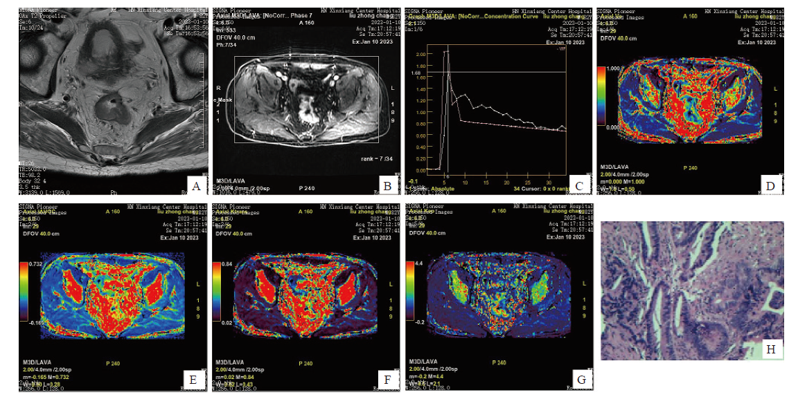
图1. DCE-MRI图像与术后病理学图像
Fig. 1. DCE-MRI image and postoperative pathology image
An 82-year-old male presented with anal distension and retraction for six months, and was subsequently diagnosed with rectal cancer by pathological examination. A: The intestinal wall in the middle and lower part of rectum is unevenly thickened, with slightly high signal on T2WI, and the lesion penetrates the muscularis propria; B: The lesion showed uneven and continuous enhancement; C: Time-signal intensity curve of DCE-MRI (pink is the lesion curve); D: Pseudo-color image, yellow-green area shows local occupation of intestinal wall; E, F: Pseudo-color image, red area shows local occupation of intestinal wall; G: Pseudo-color image, with red and yellow areas showing local occupation of intestinal wall; H: Postoperative pathological picture, diagnosed as rectal (H-E staining, ×200).