中国癌症杂志 ›› 2024, Vol. 34 ›› Issue (8): 726-733.doi: 10.19401/j.cnki.1007-3639.2024.08.002

• 论著 • 上一篇    下一篇

M1型肿瘤相关巨噬细胞在肝细胞癌组织中浸润的意义

肖锋1(), 许桐林2, 朱琳1, 肖静文1, 吴天祺3, 顾春燕1()   

  1. 1.南通市第三人民医院,南通大学附属南通第三医院病理科,江苏 南通 226006
    2.南通市第三人民医院,南通大学附属南通第三医院外科,江苏 南通 226006
    3.南通大学医学院临床医学专业,江苏 南通 226006
  • 收稿日期:2024-02-29 修回日期:2024-07-03 出版日期:2024-08-30 发布日期:2024-09-10
  • 通信作者: 顾春燕(ORCID: 0009-0007-1563-0063),硕士,主任医师,南通市第三人民医院病理科主任。
  • 作者简介:肖锋(ORCID: 0009-0009-2680-609X),硕士,副主任医师,南通市第三人民医院病理科副主任。
  • 基金资助:
    南通市市级科技计划项目(JC22022025)

Significance of infiltration of M1 tumor-associated macrophages in hepatocellular carcinoma

XIAO Feng1(), XU Tonglin2, ZHU Lin1, XIAO Jingwen1, WU Tianqi3, GU Chunyan1()   

  1. 1. Department of Pathology, the Third People's Hospital of Nantong, Affiliated Nantong Hospital Three of Nantong University, Nantong 226006, Jiangsu Province, China
    2. Department of Surgery, the Third People’s Hospital of Nantong, Affiliated Nantong Hospital Three of Nantong University, Nantong 226006, Jiangsu Province, China
    3. Clinical Medicine, Major of Medical College Nantong University, Nantong 226006, Jiangsu Province, China
  • Received:2024-02-29 Revised:2024-07-03 Published:2024-08-30 Online:2024-09-10

摘要:

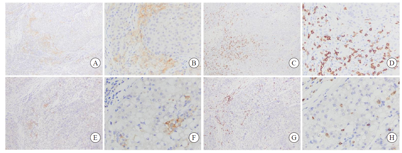
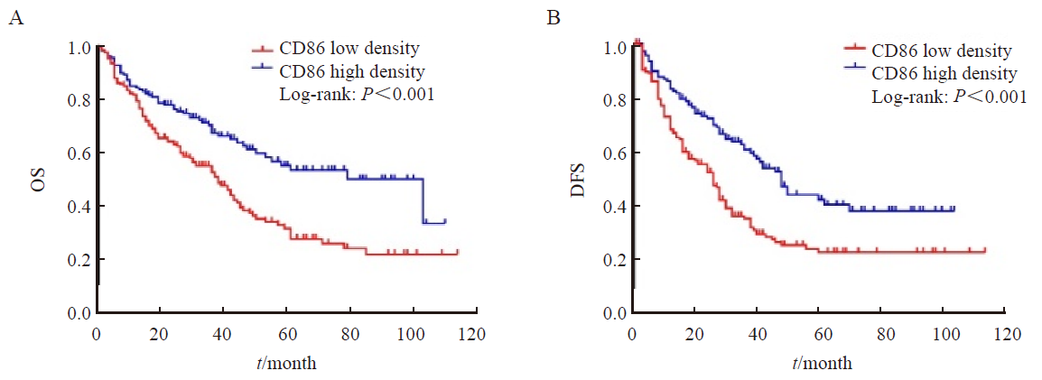
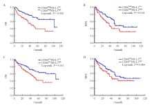
背景与目的:肿瘤相关巨噬细胞(tumor-associated macrophages,TAM)是肿瘤微环境中的主要基质细胞,在肿瘤进展过程中发挥重要作用,本研究旨在探究肝细胞癌(hepatocellular carcinoma,HCC)中M1型TAM浸润的临床意义。方法:收集2012年1月—2020年12月在南通大学附属南通第三医院接受手术的HCC患者石蜡包埋组织样本320例,采用免疫组织化学法检测CD86标记的M1型TAM在HCC组织中分布情况,计算阳性细胞密度,根据细胞密度分组:大于平均密度(29个/mm2)判定为高密度组,小于或等于平均密度为低密度组;统计分析M1型TAM密度与HCC临床病理学特征、肿瘤浸润CD8T淋巴细胞之间的相关性及预后意义;采用免疫组织化学法检测程序性死亡配体-1(programmed death ligand-1,PD-L1)的表达情况,根据CD86、PD-L1细胞密度将病例分4组:CD86高密度组中PD-L1高密度(CD86highPD-L1high)和PD-L1低密度(CD86highPD-L1low)组;CD86低密度组中PD-L1高密度(CD86lowPD-L1high)和PD-L1低密度(CD86lowPD-L1low)组,分析CD86 M1型TAM密度联合PD-L1表达的预后意义。本研究通过南通大学附属南通第三医院伦理委员会批准(伦理编号:EK2022005)。结果:CD86M1型TAM主要分布于肿瘤间质中;其高密度率为44.7%(143/320)。CD86M1型TAM密度与CD8肿瘤浸润细胞毒性T淋巴细胞密度呈正相关(P<0.001)、与乙型肝炎病毒表面抗原(hepatitis B virus surface antigen,HBsAg)阳性呈负相关(P=0.003),与患者性别、年龄、肝硬化、肿瘤大小、组织学分级、微血管侵犯等临床病理学指标均无明显相关性;CD86M1型TAM高密度组患者总生存期(overall survival,OS)、无病生存期(disease-free survival,DFS)优于低密度组,差异均有统计学意义(P均<0.001)。多因素Cox比例风险回归模型分析显示,低密度CD86M1型TAM是评估OS和DFS的独立风险因子(OS:HR=1.468,P=0.022;DFS:HR=2.233,P<0.001)。CD86highPD-L1high组HCC患者OS、DFS差于CD86highPD-L1low组,两者差异有统计学意义(P均<0.05)。CD86lowPD-L1high组OS、DFS差于CD86lowPD-L1low组,两者OS差异有统计学意义(P<0.05),DFS差异无统计学意义。结论:HCC组织中存在高密度CD86M1型TAM提示患者预后良好,并且是独立的预后因子。HCC组织表达PD-L1提示肿瘤侵袭性增强,患者预后差。

关键词: 巨噬细胞, 程序性死亡配体1, 肝细胞癌, 预后

Abstract:

Background and purpose: Tumor-associated macrophages (TAM) as the main stromal cells in the tumor microenvironment play an important role in tumor progression. This study aimed to explore the clinical significance of M1 type TAM infiltration in hepatocellular carcinoma (HCC). Methods: We collected tissue paraffin samples from 320 HCC patients who underwent surgery at the Affiliated Nantong Hospital Three of Nantong University from January 2012 to December 2020. Immunohistochemical methods were used to detect the distribution of CD86 labeled M1 type TAM in HCC tissues, and positive cell density was calculated. Groups were established according to cell density, high-density group had cells with greater than average density (29 cells/mm2), and low-density group had cells with less than or equal to average density. The correlation and prognostic significance of M1 TAM density with clinicopathologic features and tumor infiltrating CD8 T lymphocytes of HCC were analyzed. Using immunohistochemistry to detect the expression of programmed death ligand-1 (PD-L1), the cases were divided into four groups based on the cell density of CD86 and PD-L1. In the CD86 high-density group, PD-L1 high-density (CD86highPD-L1high) and PD-L1 low-density (CD86highPD-L1low) groups were included. In the CD86 low-density group, the PD-L1 high-density (CD86lowPD-L1high) and PD-L1 low-density (CD86lowPD-L1low) groups were included. We analyzed the prognostic significance of CD86 M1 type TAM density combined with PD-L1 expression. This study was approved by the Ethics Committee of Affiliated Nantong Hospital Three of Nantong University (ethics number: EK2022005). Results: CD86 M1 type TAM was mainly distributed in the tumor stroma. Its high-density rate was 44.7% (143/320). The density of CD86 M1 type TAM was positively correlated with tumor infiltrating CD8 T lymphocyte density (P<0.001) and negatively correlated with hepatitis B virus surface antigen (HBsAg) positivity (P=0.003), and had no significant correlation with clinical and pathological features such as patient age, gender, cirrhosis, tumor size, histological grading and microvascular invasion. The CD86 M1 type TAM high-density group had better overall survival (OS) and disease-free survival (DFS) than the low-density group, and the differences were statistically significant (all P<0.001). Multivariate Cox proportional hazards regression model analysis showed that low-density CD86 M1 type TAM was an independent risk factor for evaluating OS and DFS (OS: HR=1.468, P=0.022; DFS: HR=2.233, P<0.001). The CD86highPD-L1high group had poor OS and DFS than the CD86highPD-L1low group, and the differences were statistically significant (both P<0.05). The CD86lowPD-L1high group had poor OS and DFS than the CD86lowPD-L1low group. The difference in OS between the two groups was statistically significant (P<0.05), while the difference in DFS was not statistically significant. Conclusion: The presence of high-density CD86 M1 type TAM in HCC tissue suggests a good prognosis and is an independent prognostic factor. Expression of PD-L1 in HCC tissue suggests increased invasiveness and poorer prognosis.

Key words: Macrophage, Programmed death ligand 1, Hepatocellular carcinoma, Prognosis

中图分类号: