Welcome to China Oncology,
China Oncology ›› 2025, Vol. 35 ›› Issue (4): 404-411.doi: 10.19401/j.cnki.1007-3639.2025.04.008
• Article • Previous Articles Next Articles
XUE Jialei1,2( ), LI Jianwei1, GONG Yue1, LIU Guangyu1, LIU Zhebin1()
Received:2024-11-15
Revised:2025-03-24
Online:2025-04-30
Published:2025-05-16
Contact:
LIU Zhebin
Share article
XUE Jialei, LI Jianwei, GONG Yue, LIU Guangyu, LIU Zhebin. Predicting delayed diagnosis rate of intraoperative rapid frozen section pathological examination for early-stage breast cancer: a real-world retrospective study[J]. China Oncology, 2025, 35(4): 404-411.
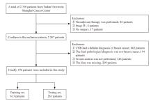
Fig. 1
Flow diagram of patients selecting"
Tab. 1
Summary sheet of the clinical data of the patients [n (%)]"
| Item | Total (n=876) | Training set (n=613) | Testing set (n=263) |
|---|---|---|---|
| Age/year | |||
| Median age | 53 (28-86) | 54 (30-86) | 51 (28-83) |
| <45 | 183 (20.9) | 127 (20.7) | 56 (21.3) |
| 45-59 | 410 (46.8) | 286 (46.6) | 124 (47.1) |
| ≥60 | 283 (32.3) | 200 (32.6) | 83 (31.6) |
| Ultrasonographic features | |||
| Echo type | |||
| Mixed echo | 82 (9.4) | 55 (9.0) | 27 (10.3) |
| Low echo | 764 (87.2) | 538 (87.8) | 226 (85.9) |
| Medium echo | 30 (3.4) | 20 (2.2) | 10 (3.8) |
| Maximum diameter D/mm | |||
| ≤10 | 111 (12.7) | 75 (12.2) | 36 (13.7) |
| >10 | 765 (87.3) | 538 (87.8) | 227 (86.3) |
| Dense punctate strong echo | |||
| Yes | 298 (34.0) | 201 (32.8) | 97 (36.9) |
| No | 578 (66.0) | 412 (67.2) | 166 (63.1) |
| BI-RADS | |||
| 1-3 | 29 (3.3) | 18 (2.9) | 11 (4.2) |
| 4A or 0 | 192 (21.9) | 136 (22.2) | 56 (21.3) |
| 4B | 344 (39.3) | 240 (39.2) | 104 (39.5) |
| 4C or 5 | 311 (35.5) | 219 (35.7) | 92 (35.0) |
| Mammographic features | |||
| Microcalcifications | |||
| Yes | 420 (47.9) | 297 (48.5) | 123 (46.8) |
| No | 456 (52.1) | 316 (51.5) | 140 (53.2) |
| BI-RADS | |||
| 1-3 | 71 (8.1) | 45 (7.3) | 26 (9.9) |
| 4A or 0 | 269 (30.7) | 182 (29.7) | 87 (33.1) |
| 4B | 247 (28.2) | 171 (27.9) | 76 (28.9) |
| 4C or 5 | 289 (33.0) | 215 (35.1) | 74 (28.1) |
| Physical examination symptoms | |||
| Palpable mass | |||
| Yes | 812 (92.7) | 571 (93.1) | 241 (91.6) |
| No | 64 (7.3) | 42 (6.9) | 22 (8.4) |
| Nipple discharge | |||
| Yes | 96 (11.0) | 69 (11.3) | 27 (10.3) |
| No | 780 (89.0) | 544 (88.7) | 236 (89.7) |
| Pathological features | |||
| Papillary lesions | |||
| Yes | 252 (28.8) | 174 (28.4) | 78 (29.7) |
| No | 624 (71.2) | 439 (71.6) | 185 (70.3) |
| Sclerosing adenosis | |||
| Yes | 61 (7.0) | 45 (7.3) | 16 (6.1) |
| No | 815 (93.0) | 568 (92.7) | 247 (93.9) |
| Frozen section | |||
| False negative | 283 (32.3) | 203 (33.1) | 80 (30.4) |
| Ture positive | 593 (67.7) | 410 (66.9) | 183 (69.6) |
Tab. 2
Results of multivariate logistic model of the training set (n=613)"
| Variables | No. of patients (delayed diagnosis/total) | DDR/% | OR | 95% CI | P value |
|---|---|---|---|---|---|
| Dense punctate strong echo | |||||
| No | 176/412 | 42.7 | 1.000 | - | - |
| Yes | 27/201 | 13.4 | 0.595 | 0.335-1.044 | 0.073 |
| US-BI-RADS | |||||
| 1-3 | 9/18 | 50.0 | 1.000 | - | - |
| 0 or 4A | 61/136 | 44.9 | 0.601 | 0.195-1.861 | 0.372 |
| 4B | 88/240 | 36.7 | 0.434 | 0.143-1.321 | 0.137 |
| 4C-5 | 45/219 | 20.5 | 0.250 | 0.081-0.777 | 0.016 |
| Microcalcifications on MG | |||||
| No | 155/316 | 49.1 | 1.000 | - | - |
| Yes | 48/297 | 16.2 | 0.345 | 0.216-0.543 | <0.001 |
| Papillary lesions | |||||
| No | 98/439 | 22.3 | 1.000 | - | - |
| Yes | 105/174 | 60.3 | 4.251 | 2.804-6.492 | <0.001 |
| Sclerosing adenosis | |||||
| No | 180/568 | 31.7 | 1.000 | - | - |
| Yes | 23/45 | 51.1 | 3.727 | 1.897-7.376 | <0.001 |
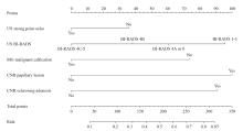
Fig. 2
Nomogram for predicting the delayed diagnosis rate of pathological diagnosis by frozen section in surgery US: Ultrosonography diagnosis."
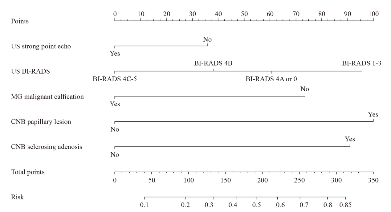
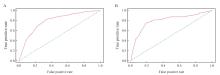
Fig. 3
ROC curve of the prediction model A: Training set with an AUC of 0.794 (95% CI: 0.756 to 0.831); B: Validation set with an AUC of 0.800 (95% CI: 0.736-0.865)."
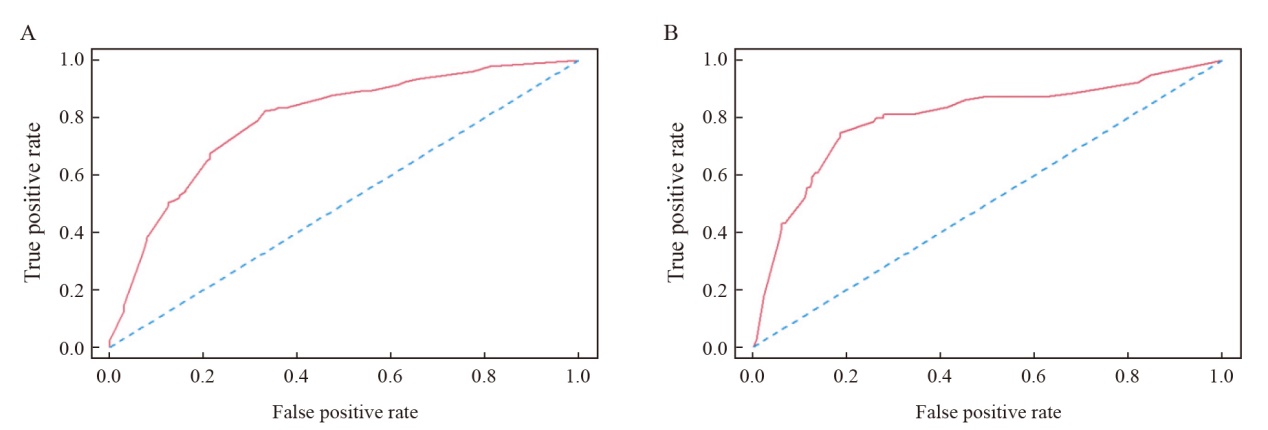
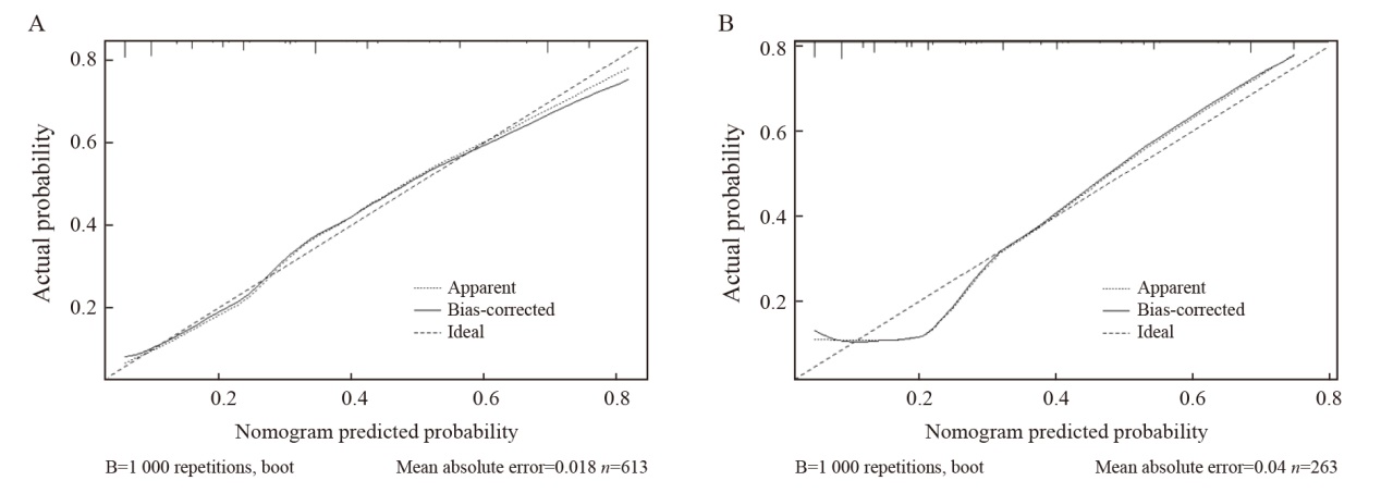
Fig. 4
Calibration curve A: Training set; B: Validation set. The horizontal axis represented the predicted DDR of the Nomogram, and the vertical axis represented the actual DDR (P=1.000)."
Tab. 3
The comparison table of predicted DDR and real DDR corresponding to different total score segments"
| Cut-off value | No. of patients n | Real DDR/% | ||
|---|---|---|---|---|
| Total score | Predicted DDR/% | Total | Delayed diagnosis | |
| ≤40 | ≤10 | 220 | 22 | 10.0 |
| ≤76 | ≤15 | 246 | 25 | 10.2 |
| ≥190 | ≥50 | 235 | 152 | 64.7 |
| ≥220 | ≥60 | 159 | 114 | 71.7 |
| ≥251 | ≥70 | 60 | 44 | 73.3 |
| ≥288 | ≥80 | 8 | 7 | 87.5 |
| [1] | MA J F, CHEN L Y, WU S L, et al. Clinical practice guidelines for ultrasound-guided breast lesions and lymph nodes biopsy: Chinese Society of Breast Surgery (CSBrS) practice guidelines 2021[J]. Chin Med J (Engl), 2021, 134(12): 1393-1395. |
| [2] | HAO S, LIU Z B, LING H, et al. Changing attitudes toward needle biopsies of breast cancer in Shanghai: experience and current status over the past 8 years[J]. Onco Targets Ther, 2015, 8: 2865-2871. |
| [3] | SCHIAFFINO S, CALABRESE M, MELANI E F, et al. Upgrade rate of percutaneously diagnosed pure atypical ductal hyperplasia: systematic review and meta-analysis of 6 458 lesions[J]. Radiology, 2020, 294(1): 76-86. |
| [4] | PAWLOSKI K R, CHRISTIAN N, KNEZEVIC A, et al. Atypical ductal hyperplasia bordering on DCIS on core biopsy is associated with higher risk of upgrade than conventional atypical ductal hyperplasia[J]. Breast Cancer Res Treat, 2020, 184(3): 873-880. |
| [5] | NIU Y, FU X L, YU Y, et al. Intra-operative frozen section diagnosis of breast lesions: a retrospective analysis of 13 243 Chinese patients[J]. Chin Med J (Engl), 2007, 120(8): 630-635. |
| [6] | 王泽英. 乳腺肿瘤冰冻切片病理诊断分析[J]. 实验与检验医学, 2023, 41(5): 669-672. |
| WANG Z Y. Pathological diagnosis of frozen section of breast tumor[J]. Exp Lab Med, 2023, 41(5): 669-672. | |
| [7] |
STOLNICU S, RĂDULESCU D, PLEŞEA I E, et al. The value of intraoperative diagnosis in breast lesions[J]. Rom J Morphol Embryol, 2006, 47(2): 119-123.
pmid: 17106518 |
| [8] | 王悦, 李晓洁, 杨璐. 探讨影响乳腺癌患者术中冰冻切片病理诊断结果准确性的临床因素[J]. 中国处方药, 2023, 21(10): 175-178. |
| WANG Y, LI X J, YANG L. To explore the clinical factors affecting the accuracy of pathological diagnosis results of intraoperative frozen section in breast cancer patients[J]. J Chin Prescr Drug, 2023, 21(10): 175-178. | |
| [9] | CSERNI G. Pitfalls in frozen section interpretation: a retrospective study of palpable breast tumors[J]. Tumori, 1999, 85(1): 15-18. |
| [10] |
FECHNER R E. Frozen section examination of breast biopsies. Practice parameter[J]. Am J Clin Pathol, 1995, 103(1): 6-7.
pmid: 7817947 |
| [11] | 施昀, 马世荣, 杨巧, 等. 手术中快速冷冻切片3 303例病理诊断分析[J]. 临床与实验病理学杂志, 2012, 28(11): 1270-1272. |
| SHI Y, MA S R, YANG Q, et al. Pathological diagnosis of 3 303 cases of quick frozen section during operation[J]. Chin J Clin Exp Pathol, 2012, 28(11): 1270-1272. | |
| [12] | 刘彤华. 不易鉴别良恶性的乳腺病变[J]. 中华病理学杂志, 1997, 26(6): 379-380. |
| LIU T H. It is difficult to distinguish benign from malignant breast lesions[J]. Chin J Pathol, 1997, 26(6): 379-380. | |
| [13] |
COUTANT C, OLIVIER C, LAMBAUDIE E, et al. Comparison of models to predict nonsentinel lymph node status in breast cancer patients with metastatic sentinel lymph nodes: a prospective multicenter study[J]. J Clin Oncol, 2009, 27(17): 2800-2808.
doi: 10.1200/JCO.2008.19.7418 pmid: 19349546 |
| [14] |
HARRELL F E Jr, LEE K L, MARK D B. Multivariable prognostic models: issues in developing models, evaluating assumptions and adequacy, and measuring and reducing errors[J]. Stat Med, 1996, 15(4): 361-387.
doi: 10.1002/(SICI)1097-0258(19960229)15:4<361::AID-SIM168>3.0.CO;2-4 pmid: 8668867 |
| [15] | STEYERBERG E W, HARRELL F E, BORSBOOM G J J M, et al. Internal validation of predictive models efficiency of some procedures for logistic regression analysis[J]. J Clin Epidemiol, 2001, 54(8): 774-781. |
| [16] | TINNEMANS J G, WOBBES T, HOLLAND R, et al. Mammographic and histopathologic correlation of nonpalpable lesions of the breast and the reliability of frozen section diagnosis[J]. Surg Gynecol Obstet, 1987, 165(6): 523-529. |
| [17] |
MOGAL H, BROWN D R, ISOM S, et al. Intracystic papillary carcinoma of the breast: a SEER database analysis of implications for therapy[J]. Breast, 2016, 27: 87-92.
doi: 10.1016/j.breast.2016.01.003 pmid: 27054753 |
| [18] | 丁雨飞, 陈建华. 乳腺实性乳头状癌临床病理研究及治疗[J]. 中华乳腺病杂志(电子版), 2014, 8(4): 280-282. |
| DING Y F, CHEN J H. Solid papillary carcinoma of the breast[J]. Chin J Breast Dis (Electro Ed), 2014, 8(4): 280-282. | |
| [19] | 曾祥菲, 魏兵. 乳腺包裹型乳头状癌的病理诊断[J]. 临床与实验病理学杂志, 2024, 40(9): 901-906. |
| ZENG X F, WEI B. Pathological diagnosis of encapsulated papillary carcinoma of breast[J]. Chin J Clin Exp Pathol, 2024, 40(9): 901-906. | |
| [20] |
郑小草, 葛荣, 蒙伶俐, 等. 乳腺实性乳头状癌的临床病理研究[J]. 中国癌症杂志, 2014, 24(3): 208-211.
doi: 10.3969/j.issn.1007-3969.2014.03.009 |
| ZHENG X C, GE R, MENG L L, et al. Clinicopathologic study of solid papillary carcinoma of the breast[J]. Chin Oncol, 2014, 24(3): 208-211. | |
| [21] |
TAN P H, SCHNITT S J, VAN DE VIJVER M J, et al. Papillary and neuroendocrine breast lesions: the WHO stance[J]. Histopathology, 2015, 66(6): 761-770.
doi: 10.1111/his.12463 pmid: 24845113 |
| [22] |
WANG H, TSANG P, D’CRUZ C, et al. Follow-up of breast papillary lesion on core needle biopsy: experience in African-American population[J]. Diagn Pathol, 2014, 9: 86.
doi: 10.1186/1746-1596-9-86 pmid: 24762090 |
| [23] | YU B H, TANG S X, XU X L, et al. Breast carcinoma in sclerosing adenosis: a clinicopathological and immunophenotypical analysis on 206 lesions[J]. J Clin Pathol, 2018, 71(6): 546-553. |
| [24] | RICHARDS D, AYALA A A, WU Y, et al. Carcinoma in situ involving sclerosing adenosis on core biopsy: diagnostic pearls to aid the practicing clinician and avoid overtreatment[J]. Oncol Ther, 2020, 8(1): 81-89. |
| [25] | 卢佳, 胡兵. 误诊乳腺广泛导管内乳头状瘤合并硬化性腺病为乳腺癌1例[J]. 中国介入影像与治疗学, 2022, 19(9): 607. |
| LU J, HU B. Extensive intraductal papilloma of breast with sclerosing adenosis as breast cancer: case report[J]. Chin J Interv Imag Ther, 2022, 19(9): 607. | |
| [26] | EUSEBI V, COLLINA G, BUSSOLATI G. Carcinoma in situ in sclerosing adenosis of the breast: an immunocytochemical study[J]. Semin Diagn Pathol, 1989, 6(2): 146-152. |
| [27] | 付丽梅, 孙向洁, 吕泓, 等. 乳腺硬化性腺病基础上的低级别腺鳞癌1例[J]. 中华病理学杂志, 2019, 48(5): 415-417. |
| FU L M, SUN X J, LYU H, et al. Low grade adenosquamous carcinoma arising from sclerosing adenosis of the breast: report of a case[J]. Chin J Pathol, 2019, 48(5): 415-417. | |
| [28] | 张久存, 马爱玲. 13例乳腺硬化性腺病临床病理分析[J]. 宁夏医科大学学报, 2010, 32(1): 134-135. |
| ZHANG J C, MA A L. Clinicopathological analysis of 13 cases of breast sclerosing gonadism[J]. J Ningxia Med Univ, 2010, 32(1): 134-135. | |
| [29] | 皋岚湘, 丁华野, 杨光之, 等. 乳腺粗针穿刺活检219例病理诊断分析、问题及对策[J]. 诊断病理学杂志, 2009, 16(3): 166-170. |
| GAO L X, DING H Y, YANG G Z, et al. Problems and strategy in pathologic diagnosis of mammary needle core biopsies: an analysis of 219 cases[J]. Chin J Diagn Pathol, 2009, 16(3): 166-170. | |
| [30] | CUI X Y, WEI S. Carcinoma in situ involving sclerosing adenosis: seeking the salient histological characteristics to prevent overdiagnosis[J]. Ann Clin Lab Sci, 2017, 47(5): 529-534. |
| [31] | LI S J, HAO X P, HUA B, et al. Clinical practice guidelines for ultrasound-guided vacuum-assisted breast biopsy: Chinese Society of Breast Surgery (CSBrS) practice guidelines 2021[J]. Chin Med J (Engl), 2021, 134(12): 1390-1392. |
| Viewed | ||||||
|
Full text |
|
|||||
|
Abstract |
|
|||||
沪ICP备12009617
Powered by Beijing Magtech Co. Ltd