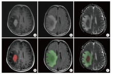
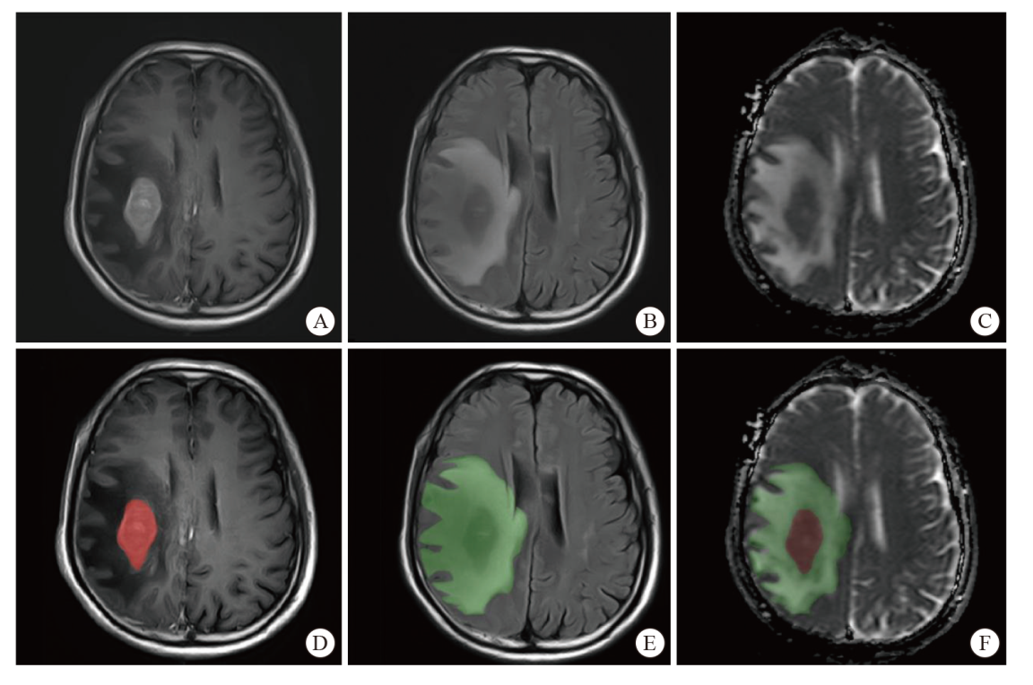
| T1-CE+T2-FLAIR+ADC | SVM | 0.887 (0.789-0.984)/0.833/0.778 | 0.822 (0.700-0.944)/0.889/0.833 |
| LR | 0.878 (0.771-0.987)/0.854/0.800 | 0.840 (0.718-0.960)/0.833/0.769 |
| GP | 0.900 (0.805-0.906)/0.896/0.840 | 0.834 (0.710-0.958)/0.889/0.833 |
| NB | 0.805 (0.678-0.932)/0.813/0.743 | 0.852 (0.738-0.965)/0.833/0.800 |
| DenseNet121 | 0.846 (0.710-0.982)/0.854/0.774 | 0.807 (0.672-0.941)/0.889/0.875 |
| ResNet101 | 0.842 (0.701-0.983)/0.833/0.750 | 0.826 (0.706-0.947)/0.833/0.800 |
| EfficientNet-b5 | 0.802 (0.674-0.929)/0.771/0.703 | 0.802 (0.673-0.937)/0.889/0.833 |
| T1-CE+T2-FLAIR | SVM | 0.709 (0.562-0.856)/0.646/0.622 | 0.703 (0.552-0.854)/0.722/0.615 |
| LR | 0.719 (0.572-0.866)/0.667/0.636 | 0.703 (0.552-0.854)/0.778/0.714 |
| GP | 0.719 (0.574-0.863)/0.667/0.652 | 0.678 (0.517-0.839)/0.611/0.588 |
| NB | 0.688 (0.529-0.846)/0.708/0.632 | 0.717 (0.571-0.863)/0.556/0.556 |
| DenseNet121 | 0.713 (0.565-0.860)/0.646/0.622 | 0.691 (0.526-0.856)/0.667/0.666 |
| ResNet101 | 0.723 (0.576-0.869)/0.729/0.629 | 0.738 (0.585-0.892)/0.611/0.632 |
| EfficientNet-b5 | 0.725 (0.582-0.867)/0.688/0.651 | 0.732 (0.578-0.886)/0.611/0.364 |
| T1-CE+ADC | SVM | 0.793 (0.660-0.926)/0.750/0.700 | 0.562 (0.375-0.750)/0.778/0.666 |
| LR | 0.840 (0.718-0.962)/0.771/0.718 | 0.615 (0.440-0.790)/0.778/0.714 |
| GP | 0.844 (0.726-0.961)/0.792/0.750 | 0.547 (0.359-0.734)/0.611/0.588 |
| NB | 0.820 (0.696-0.945)/0.792/0.722 | 0.572 (0.394-0.750)/0.556/0.556 |
| DenseNet121 | 0.822 (0.700-0.944)/0.771/0.718 | 0.531 (0.344-0.718)/0.778/0.666 |
| ResNet101 | 0.840 (0.718-0.962)/0.771/0.718 | 0.578 (0.396-0.760)/0.611/0.364 |
| EfficientNet-b5 | 0.852 (0.738-0.965)/0.771/0.732 | 0.611 (0.438-0.785)/0.667/0.625 |
| T2-FLAIR+ADC | SVM | 0.822 (0.700-0.944)/0.771/0.718 | 0.709 (0.546-0.872)/0.722/0.635 |
| LR | 0.840 (0.718-0.962)/0.771/0.718 | 0.537 (0.359-0.716)/0.667/0.571 |
| GP | 0.852 (0.738-0.965)/0.771/0.732 | 0.525 (0.336-0.715)/0.611/0.588 |
| NB | 0.834 (0.710-0.958)/0.833/0.765 | 0.740 (0.585-0.895)/0.778/0.714 |
| DenseNet121 | 0.834 (0.710-0.958)/0.833/0.765 | 0.568 (0.392-0.745)/0.833/0.800 |
| ResNet101 | 0.793 (0.660-0.926)/0.750/0.700 | 0.525 (0.339-0.711)/0.722/0.615 |
| EfficientNet-b5 | 0.844 (0.722-0.966)/0.771/0.718 | 0.570 (0.388-0.753)/0.722/0.615 |
| T1-CE | SVM | 0.527 (0.338-0.717)/0.667/0.385 | 0.527 (0.338-0.717)/0.444/0.445 |
| LR | 0.562 (0.377-0.748)/0.625/0.471 | 0.562 (0.377-0.748)/0.500/0.470 |
| GP | 0.549 (0.367-0.730)/0.646/0.452 | 0.529 (0.342-0.717)/0.556/0.556 |
| NB | 0.529 (0.342-0.717)/0.667/0.385 | 0.549 (0.367-0.730)/0.444/0.375 |
| DenseNet121 | 0.721 (0.565-0.876)/0.708/0.632 | 0.721 (0.565-0.876)/0.611/0.364 |
| ResNet101 | 0.705 (0.541-0.869)/0.729/0.552 | 0.705 (0.542-0.869)/0.444/0.375 |
| EfficientNet-b5 | 0.684 (0.516-0.851)/0.708/0.611 | 0.684 (0.517-0.852)/0.667/0.625 |
| T2-FLAIR | SVM | 0.703 (0.552-0.854)/0.708/0.611 | 0.518 (0.329-0.708)/0.722/0.666 |
| LR | 0.703 (0.552-0.854)/0.688/0.615 | 0.562 (0.377-0.748)/0.778/0.714 |
| GP | 0.717 (0.571-0.863)/0.667/0.619 | 0.525 (0.336-0.715)/0.611/0.588 |
| NB | 0.678 (0.517-0.839)/0.729/0.587 | 0.553 (0.372-0.733)/0.722/0.666 |
| DenseNet121 | 0.711 (0.563-0.859)/0.708/0.611 | 0.611 (0.438-0.785)/0.722/0.615 |
| ResNet101 | 0.703 (0.552-0.854)/0.688/0.615 | 0.539 (0.365-0.713)/0.667/0.625 |
| EfficientNet-b5 | 0.711 (0.560-0.861)/0.729/0.606 | 0.539 (0.366-0.713)/0.667/0.666 |
| ADC | SVM | 0.779 (0.622-0.936)/0.750/0.690 | 0.713 (0.565-0.860)/0.778/0.667 |
| LR | 0.807 (0.666-0.947)/0.833/0.661 | 0.723 (0.576-0.869)/0.833/0.769 |
| GP | 0.836 (0.712-0.959)/0.750/0.714 | 0.691 (0.536-0.847)/0.667/0.625 |
| NB | 0.819 (0.690-0.948)/0.833/0.734 | 0.725 (0.582-0.867)/0.556/0.556 |
| DenseNet121 | 0.807 (0.674-0.934)/0.771/0.703 | 0.723 (0.563-0.882)/0.833/0.800 |
| ResNet101 | 0.770 (0.614-0.925)/0.729/0.683 | 0.691 (0.528-0.855)/0.833/0.769 |
| EfficientNet-b5 | 0.834 (0.714-0.954)/0.792/0.706 | 0.719 (0.559-0.879)/0.722/0.706 |
 ), 徐智坚4, 曹鑫1,2,3, 吕锟1,2,3, 李惠明5, 高敏6, 居胜红5, 刘军6, 耿道颖1,2,3(