中国癌症杂志 ›› 2024, Vol. 34 ›› Issue (4): 389-399.doi: 10.19401/j.cnki.1007-3639.2024.04.006

• 论著 • 上一篇    下一篇

HMGA2对软脑膜转移黑色素瘤细胞迁移和增殖的影响

李晓辉1(), 赵加旭1, 彭海豹1, 张叶1, 曾睿1,2, 迟喻丹1,2()   

  1. 1.复旦大学脑科学转化研究院,脑功能与脑疾病全国重点实验室,脑科学前沿科学中心,上海 200032
    2.复旦大学附属华山医院神经外科,上海 200040
  • 收稿日期:2023-09-21 修回日期:2023-12-27 出版日期:2024-04-30 发布日期:2024-05-17
  • 通信作者: 迟喻丹(ORCID: 0000-0002-4566-3305),研究员。
  • 作者简介:李晓辉(ORCID: 0009-0006-2715-9427),硕士研究生在读。
  • 基金资助:
    国家自然科学基金(32200640)

Effects of HMGA2 on migration and proliferation of leptomeningeal metastatic melanoma

LI Xiaohui1(), ZHAO Jiaxu1, PENG Haibao1, ZHANG Ye1, ZENG Rui1,2, CHI Yudan1,2()   

  1. 1. Institute for Translational Brain Research, State Key Laboratory of Medical Neurobiology, MOE Frontiers Center for Brain Science, Fudan University, Shanghai 200032, China
    2. Department of Neurosurgery, Huashan Hospital, Fudan University, Shanghai 200040, China
  • Received:2023-09-21 Revised:2023-12-27 Published:2024-04-30 Online:2024-05-17
  • Contact: CHI Yudan

摘要:

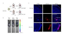
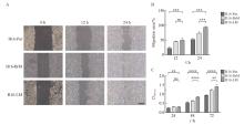
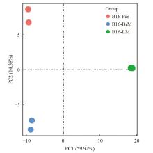
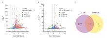
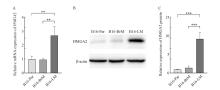
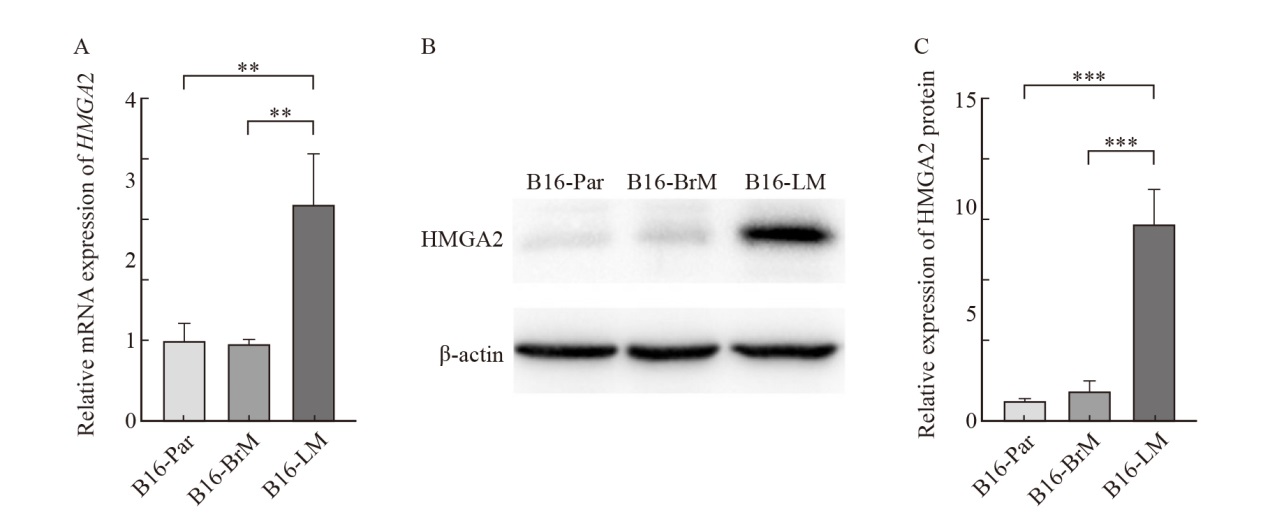
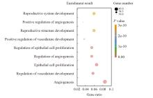
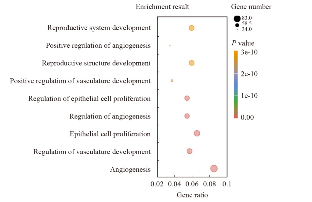
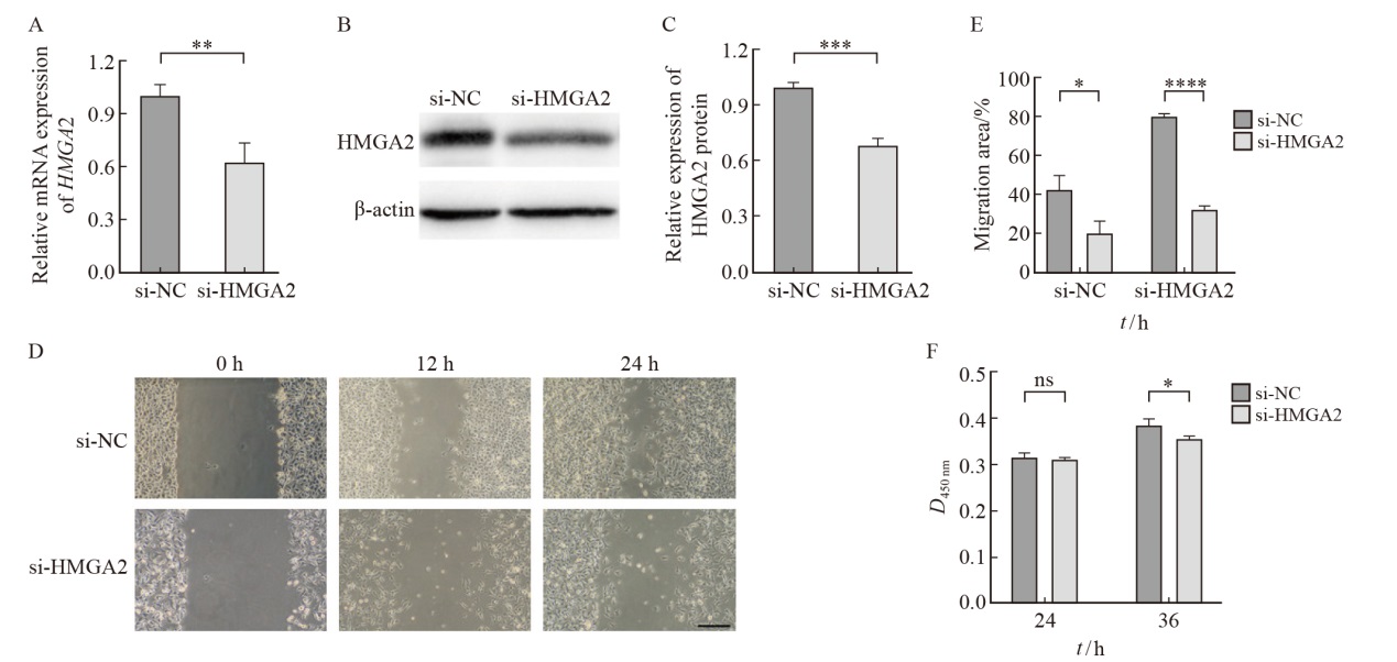
背景与目的:软脑膜转移是黑色素瘤中枢神经系统转移的一种形式,高迁移率族蛋白A2(high-mobility group protein A2,HMGA2)已被证实在多种肿瘤的发生、发展过程中起重要作用,但在软脑膜转移黑色素瘤细胞中的生物学作用尚未明确。本研究在构建黑色素瘤中枢神经系统转移小鼠模型的基础上,探讨软脑膜转移黑色素瘤与原发部位以及脑实质转移黑色素瘤在细胞迁移、细胞增殖等方面的差异,并进一步明确差异表达基因HMGA2对软脑膜转移黑色素瘤细胞迁移和增殖的影响。方法:通过慢病毒感染构建稳定表达tdTomato和荧光素酶的B16小鼠黑色素瘤细胞(B16-parental cell,B16-Par)。在此基础上,经过体内转移部位适应性筛选,获得B16特异性脑实质转移细胞(B16-brain metastatic cell,B16-BrM)以及B16特异性软脑膜转移细胞(B16-leptomeningeal metastatic cell,B16-LM)。采用细胞划痕实验以及细胞计数试剂盒-8(cell counting kit-8,CCK-8)检测B16-Par、B16-BrM和B16-LM在迁移和增殖能力上的差异。利用转录组测序技术(RNA sequencing,RNA-seq)分析B16-Par、B16-BrM和B16-LM的差异基因表达,筛选出在B16-LM中特异性上调的HMGA2基因,用实时荧光定量聚合酶链反应(real-time fluorescence quantitative polymerase chain reaction,RTFQ-PCR)和蛋白质印迹法(Western blot)对该结果进行验证。对B16-LM特异性上调基因进行基因本体论(Gene Ontology,GO)分析。使用siRNA干扰HMGA2基因在B16-LM中的表达,通过RTFQ-PCR和Western blot实验对敲低效果进行验证。细胞划痕实验和CCK-8实验检测敲低HMGA2对细胞迁移和增殖的影响。利用基因表达综合数据库(Gene Expression Omnibus,GEO)中GSE174401的数据,验证HMGA2基因在患者软脑膜转移黑色素瘤细胞中表达的特异性。结果:与B16-Par相比,经过脑内环境筛选的肿瘤细胞更易于在中枢神经系统定植。B16-LM具有更强的迁移和增殖能力,且上调表达HMGA2基因。GO分析显示,HMGA2与血管生成、细胞增殖等多个生物学过程相关。敲低HMGA2基因的表达可以抑制B16-LM的迁移和增殖。HMGA2在患者软脑膜转移黑色素瘤细胞中表达上调。结论:软脑膜转移黑色素瘤细胞具有相对独特的细胞生物学行为,其通过上调HMGA2基因的表达来促进细胞迁移和增殖。

关键词: 黑色素瘤, 中枢神经系统转移, 软脑膜转移, 脑转移, HMGA2

Abstract:

Background and purpose: Leptomeningeal metastasis is a form of central nervous system metastasis of melanoma. High mobility group A2 (HMGA2) has been proven to play an important role in the occurrence and development of various tumors, but its biological functions in leptomeningeal metastatic melanoma cells remain unclear. On the basis of building mouse models of central nervous system metastasis of melanoma, this study investigated the differences in cell migration and cell proliferation among leptomeningeal metastatic melanoma cells, primary site melanoma cells and brain parenchymal metastatic melanoma cells, and further clarified the effects of differentially expressed gene HMGA2 on cell migration and proliferation of leptomeningeal metastatic melanoma cells. Methods: B16 mouse melanoma cells (B16-parental cells, B16-Par) stably expressing tdTomato and luciferase were generated by lentiviral infection. Subsequently, B16 specific brain parenchymal metastatic cells (B16-brain metastatic cells, B16-BrM) and B16 specific leptomeningeal metastatic cells (B16-leptomeningeal metastatic cells, B16-LM) were collected after adaptive screening of metastatic sites in vivo. The differences in migration and proliferation among B16-Par, B16-BrM and B16-LM were assessed by wound healing assay and cell counting kit-8 (CCK-8). RNA sequencing (RNA-seq) was used to analyze differential gene expression in B16-Par, B16-BrM and B16-LM, and HMGA2 gene specifically upregulated in B16-LM was screened out. The results were verified by real-time fluorescence quantitative polymerase chain reaction (RTFQ-PCR) and Western blot. Gene ontology (GO) analysis was performed for genes which were upregulated in B16-LM specifically. siRNA was used to interfere with the expression of HMGA2 gene in B16-LM, and the knock-down effect was verified by RTFQ-PCR and Western blot. The effects of knocking down HMGA2 on cell migration and proliferation were detected by wound healing assay and CCK-8 assay. Using GSE174401 data in Gene Expression Omnibus (GEO), the specificity of HMGA2 gene expression in leptomeningeal metastatic melanoma cells from patients was verified. Results: Compared with Par cells, tumor cells screened by the brain environment were more likely to colonize the central nervous system. B16-LM had stronger migration and proliferation abilities, and upregulated the expression of HMGA2 gene. GO analysis revealed that HMGA2 was associated with many biological processes such as angiogenesis and cell proliferation. When the expression of HMGA2 gene was knocked down, the migration and proliferation of B16-LM could be inhibited. HMGA2 was upregulated in leptomeningeal metastatic melanoma cells from patients. Conclusion: Leptomeningeal metastatic melanoma cells had relatively unique cellular characteristics, which promoted cell migration and proliferation by upregulating HMGA2 gene expression.

Key words: Melanoma, Central nervous system metastasis, Leptomeningeal metastasis, Brain metastasis, HMGA2

中图分类号: