Welcome to China Oncology,
China Oncology ›› 2025, Vol. 35 ›› Issue (9): 859-866.doi: 10.19401/j.cnki.1007-3639.2025.09.006
• Article • Previous Articles Next Articles
WANG Tong( ), SUN Wei, XU Yu, HU Tu, LIU Wanlin, ZHENG Qiongdan, ZOU Zijian, DONG Zirui, MA Wenjie, CHEN Yong()
Received:2025-05-21
Revised:2025-07-15
Online:2025-09-30
Published:2025-10-17
Contact:
CHEN Yong
Supported by:Share article
CLC Number:
WANG Tong, SUN Wei, XU Yu, HU Tu, LIU Wanlin, ZHENG Qiongdan, ZOU Zijian, DONG Zirui, MA Wenjie, CHEN Yong. MITF expression in acral melanoma tissues and its association with clinical, pathological characteristics and prognosis[J]. China Oncology, 2025, 35(9): 859-866.
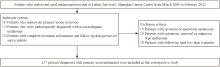
Fig. 1
Flowchart of patient inclusion and exclusion criteria"
Tab. 1
Basic characteristics, clinical, pathological features and adjuvant therapeutic schedules of study participants stratified by MITF expression levels [n (%)]"
| Characteristic | MITF-low (n=82) | MITF-high (n=55) | Pearson χ2 | P value | Characteristic | MITF-low (n=82) | MITF-high (n=55) | Pearson χ2 | P value | |
|---|---|---|---|---|---|---|---|---|---|---|
| Gender | 0.029 | 0.864 | Ⅳ | 52 (63.41) | 27 (49.09) | |||||
| Male | 45 (54.88) | 31 (56.36) | Ⅴ | 13 (15.85) | 21 (38.18) | |||||
| Female | 37 (45.12) | 24 (43.64) | Unknown | 5 (6.10) | 3 (5.45) | |||||
| Age at diagnosis/year | 3.387 | 0.066 | SLN | 10.793 | 0.001 | |||||
| ≤60 | 26 (31.71) | 26 (47.27) | Positive | 16 (19.51) | 20 (36.36) | |||||
| >60 | 56 (68.29) | 29 (52.73) | Negative | 50 (60.98) | 15 (27.27) | |||||
| T stage | 9.459 | 0.024 | Unknown | 16 (19.51) | 20 (36.36) | |||||
| T1 | 11 (13.41) | 1 (1.82) | Ulceration | 7.583 | 0.006 | |||||
| T2 | 14 (17.07) | 5 (9.09) | Positive | 34 (41.46) | 36 (65.45) | |||||
| T3 | 27 (32.93) | 18 (32.73) | Negative | 48 (58.54) | 19 (34.55) | |||||
| T4 | 30 (36.59) | 31 (56.36) | Location | 0.532 | 0.466 | |||||
| N stage | 21.937 | <0.001 | Upper extremity | 6 (7.32) | 6 (10.91) | |||||
| N0 | 55 (67.07) | 16 (29.09) | Lower extremity | 76 (92.68) | 49 (89.09) | |||||
| N1 | 13 (15.85) | 14 (25.45) | Histology | 2.198 | 0.532 | |||||
| N2 | 10 (12.20) | 12 (21.82) | ALM | 52 (63.41) | 34 (61.82) | |||||
| N3 | 4 (4.88) | 13 (23.64) | NM | 3 (3.66) | 4 (7.27) | |||||
| AJCC Stage | 21.42 | <0.001 | SSM | 2 (2.44) | 0 (0.00) | |||||
| Ⅰ | 20 (24.39) | 2 (3.64) | Unknown | 25 (30.49) | 17 (30.91) | |||||
| Ⅱ | 35 (42.68) | 14 (25.45) | Adjuvant therapy | 12.463 | 0.006 | |||||
| Ⅲ | 27 (32.93) | 39 (70.91) | PD-1 inhibitors | 22 (26.83) | 27 (49.09) | |||||
| Clark level | 12.531 | 0.006 | Cytokines | 29 (35.37) | 6 (10.91) | |||||
| Ⅱ | 7 (8.54) | 0 (0.00) | Observation | 24 (29.27) | 18 (32.73) | |||||
| Ⅲ | 5 (6.10) | 4 (7.27) | Unknown | 7 (8.54) | 4 (7.27) |
Tab. 2
Univariate and multivariate analysis of MITF expression level"
| Characteristic | Univariate analysis | Multivariate analysis | |||||
|---|---|---|---|---|---|---|---|
| OR | 95% CI | P value | OR | 95% CI | P value | ||
| Gender | 1.062 | 0.534-2.113 | 0.864 | ||||
| Age at diagnosis | 0.518 | 0.256-1.048 | 0.067 | ||||
| T stage | 1.872 | 1.234-2.841 | 0.003 | 1.116 | 0.618-2.018 | 0.716 | |
| N stage | 2.168 | 1.520-3.096 | <0.001 | 1.893 | 1.281-2.795 | 0.001 | |
| AJCC Stage | 4.183 | 2.040-6.746 | <0.001 | ||||
| Clark level | 2.418 | 1.336-4.380 | 0.004 | 1.473 | 0.703-3.083 | 0.305 | |
| SLN | 4.166 | 1.737-9.994 | 0.001 | ||||
| Ulceration | 2.901 | 1.420-5.930 | 0.003 | 2.907 | 1.239-6.814 | 0.014 | |
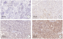
Fig. 2
MITF stained by immunohistochemistry in AM tissues A: Negative; B: Low positive; C: Positive; D: High positive."
Tab. 3
Analysis of prognostic factors in AM patients"
| Characteristic | Univariate analysis | Multivariate analysis | |||||
|---|---|---|---|---|---|---|---|
| HR | 95% CI | P value | HR | 95% CI | P value | ||
| MITF by IHC | 1.844 | 1.048-3.246 | 0.034 | 2.415 | 1.093-5.337 | 0.029 | |
| Gender | 1.991 | 1.120-3.539 | 0.019 | 1.750 | 0.773-3.961 | 0.179 | |
| Age at diagnosis | 0.784 | 0.453-1.358 | 0.389 | ||||
| T stage | 1.777 | 1.237-2.553 | 0.002 | 1.974 | 1.083-3.599 | 0.026 | |
| N stage | 1.195 | 0.930-1.534 | 0.163 | ||||
| Clark level | 1.416 | 0.886-2.264 | 0.146 | 0.854 | 0.403-1.807 | 0.679 | |
| SLN | 1.390 | 0.713-2.710 | 0.333 | 1.503 | 0.700-3.226 | 0.296 | |
| Ulceration | 0.630 | 0.663-1.972 | 0.630 | ||||
| Adjuvant therapy | 1.261 | 0.704-2.261 | 0.436 | 1.253 | 0.565-2.776 | 0.579 | |
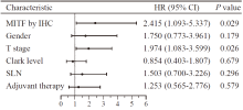
Fig. 3
Forest plot for analysis of prognostic factors in AM patients"
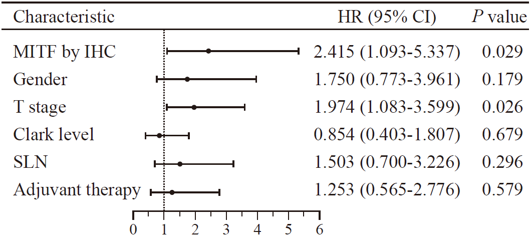
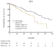
Fig. 4
DFS for patients with AM stratified based on MITF expression levels"
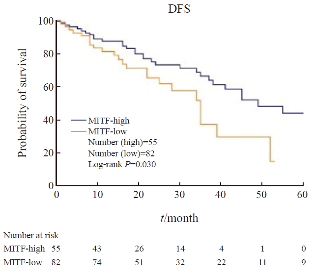
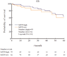
Fig. 5
OS for patients with AM stratified based on MITF expression levels"
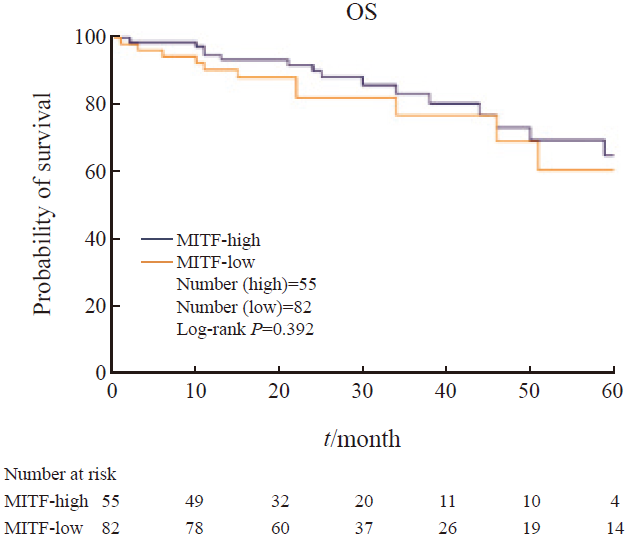
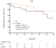
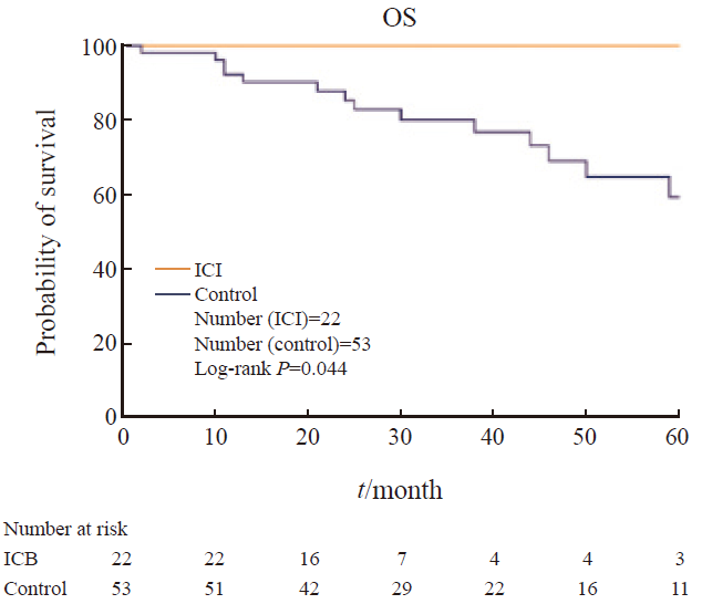
Fig. 6
Effect of ICI on OS in patients with high MITF expression"
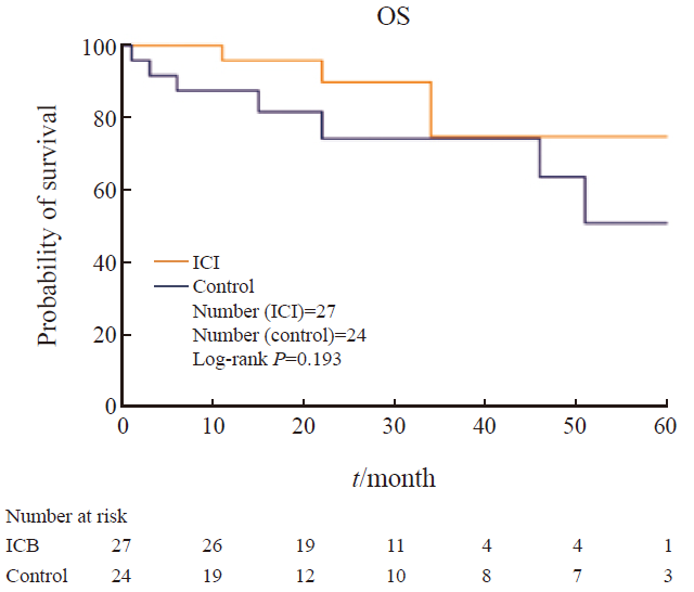
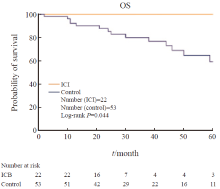
Fig. 7
Effect of ICI on OS in patients with low MITF expression"
| [1] |
CHEN Y A, TEER J K, EROGLU Z, et al. Translational pathology, genomics and the development of systemic therapies for acral melanoma[J]. Semin Cancer Biol, 2020, 61: 149-157.
doi: S1044-579X(19)30343-8 pmid: 31689494 |
| [2] | CHI Z H, LI S M, SHENG X N, et al. Clinical presentation, histology, and prognoses of malignant melanoma in ethnic Chinese: a study of 522 consecutive cases[J]. BMC Cancer, 2011, 11(1): 85. |
| [3] |
HARTMAN M L, CZYZ M. MITF in melanoma: mechanisms behind its expression and activity[J]. Cell Mol Life Sci, 2015, 72(7): 1249-1260.
doi: 10.1007/s00018-014-1791-0 pmid: 25433395 |
| [4] | HEALTH CHINA N. National guidelines for diagnosis and treatment of melanoma 2022 in China (English version)[J]. Chin J Cancer Res, 2022, 34(4): 335-342. |
| [5] |
邹孜瑊, 孙伟, 胡涂, 等. 前哨淋巴结活检在皮肤型和肢端型黑色素瘤临床诊疗中的价值[J]. 中国癌症杂志, 2022, 32(12): 1168-1177.
doi: 10.19401/j.cnki.1007-3639.2022.12.004 |
| ZOU Z J, SUN W, HU T, et al. Clinical value of sentinel lymph node biopsy in patients with cutaneous and acral melanoma[J]. China Oncol, 2022, 32(12): 1168-1177. | |
| [6] | NEWELL F, WILMOTT J S, JOHANSSON P A, et al. Whole-genome sequencing of acral melanoma reveals genomic complexity and diversity[J]. Nat Commun, 2020, 11(1): 5259. |
| [7] |
陈柳含, 张洋洋, 李先安. 肢端恶性黑色素瘤临床特征及预后因素分析[J]. 中国癌症杂志, 2019, 29(5): 362-371.
doi: 10.19401/j.cnki.1007-3639.2019.05.006 |
| CHEN L H, ZHANG Y Y, LI X A. Analysis of clinical characteristics and prognostic factors of acral melanoma[J]. China Oncol, 2019, 29(5): 362-371. | |
| [8] | KLEMEN N D, WANG M, RUBINSTEIN J C, et al. Survival after checkpoint inhibitors for metastatic acral, mucosal and uveal melanoma[J]. J Immunother Cancer, 2020, 8(1): e000341. |
| [9] | CARVALHO L A D, AGUIAR F C, SMALLEY K S M, et al. Acral melanoma: new insights into the immune and genomic landscape[J]. Neoplasia, 2023, 46: 100947. |
| [10] | GARRAWAY L A, WIDLUND H R, RUBIN M A, et al. Integrative genomic analyses identify MITF as a lineage survival oncogene amplified in malignant melanoma[J]. Nature, 2005, 436(7047): 117-122. |
| [11] | KUMAR S M, DAI J, LI S, et al. Human skin neural crest progenitor cells are susceptible to BRAF (V600E)-induced transformation[J]. Oncogene, 2014, 33(7): 832-841. |
| [12] | RAMBOW F, MARINE J C, GODING C R. Melanoma plasticity and phenotypic diversity: therapeutic barriers and opportunities[J]. Genes Dev, 2019, 33(19/20): 1295-1318. |
| [13] |
GIULIANO S, CHELI Y, OHANNA M, et al. Microphthalmia-associated transcription factor controls the DNA damage response and a lineage-specific senescence program in melanomas[J]. Cancer Res, 2010, 70(9): 3813-3822.
doi: 10.1158/0008-5472.CAN-09-2913 pmid: 20388797 |
| [14] | AUGUSTIN R C, NEWMAN S, LI A F, et al. Identification of tumor-intrinsic drivers of immune exclusion in acral melanoma[J]. J Immunother Cancer, 2023, 11(10): e007567. |
| [15] | QUEK C, PRATAPA A, BAI X Y, et al. Single-cell spatial multiomics reveals tumor microenvironment vulnerabilities in cancer resistance to immunotherapy[J]. Cell Rep, 2024, 43(7): 114392. |
| [16] | VIVAS-GARCÍA Y, FALLETTA P, LIEBING J, et al. Lineage-restricted regulation of SCD and fatty acid saturation by MITF controls melanoma phenotypic plasticity[J]. Mol Cell, 2020, 77(1): 120-137.e9. |
| [17] | MALISSEN N, MACAGNO N, GRANJEAUD S, et al. HVEM has a broader expression than PD-L1 and constitutes a negative prognostic marker and potential treatment target for melanoma[J]. Oncoimmunology, 2019, 8(12): e1665976. |
| [18] |
LIU Z Z, CHEN K G, DAI J, et al. A unique hyperdynamic dimer interface permits small molecule perturbation of the melanoma oncoprotein MITF for melanoma therapy[J]. Cell Res, 2023, 33(1): 55-70.
doi: 10.1038/s41422-022-00744-5 pmid: 36588115 |
| [19] | WEI C Y, SUN W, SHEN K J, et al. Delineating the early dissemination mechanisms of acral melanoma by integrating single-cell and spatial transcriptomic analyses[J]. Nat Commun, 2023, 14(1): 8119. |
| [20] | LUKE J J, RUTKOWSKI P, QUEIROLO P, et al. Pembrolizumab versus placebo as adjuvant therapy in completely resected stage ⅡB or ⅡC melanoma (KEYNOTE-716): a randomised, double-blind, phase 3 trial[J]. Lancet, 2022, 399(10336): 1718-1729. |
| Viewed | ||||||
|
Full text |
|
|||||
|
Abstract |
|
|||||
沪ICP备12009617
Powered by Beijing Magtech Co. Ltd