Welcome to China Oncology,
|
||
|
Expression and effect of long non-coding RNA ARAP1-AS1 in clear cell renal cell carcinoma
China Oncology
2022, 32 (1):
34-40.
DOI: 10.19401/j.cnki.1007-3639.2022.01.004
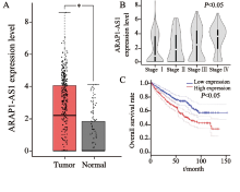
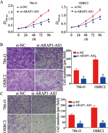
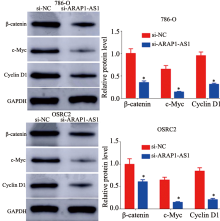
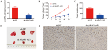
Background and purpose: Long non-coding RNA (lncRNA) ARAP1-AS1 is abnormally expressed in a variety of tumors, but its role in clear cell renal cell carcinoma (ccRCC) is unknown. This study aimed to explore the biological role of ARAP1-AS1 in ccRCC. Methods: The expression level of ARAP1-AS1 in ccRCC tissues and its relationship to patient’s clinicopathological characteristics and survival rate were analyzed using the GEPIA database. The expression level of ARAP1-AS1 was measured in ccRCC and adjacent non-tumor tissues by real-time fluorescence quantitative polymerase chain reaction (RTFQ-PCR). Patients were divided into ARAP1-AS1 high and low expression groups, the relationship between ARAP1-AS1 expression level and patient’s clinicopathological characteristics was analyzed, and survival analysis was performed. The effect of ARAP1-AS1 in vitro proliferation, migration and invasion ability of ccRCC cells was determined by cell counting kit-8 (CCK-8) assay, transwell migration and invasion assay. Changes in the expressions of Wnt/β-catenin signaling pathway-related proteins were detected by Western blot assay. The effect of ARAP1-AS1 on tumorigenic capacity in ccRCC cells in vivo was verified by tumor xenografts in nude mice. Results: The analysis of the GEPIA database showed that ARAP1-AS1 was highly expressed in ccRCC and associated with advanced tumor stage as well as poor survival in patients (all P<0.05). The results of RTFQ-PCR showed that ARAP1-AS1 was highly expressed in ccRCC tissues and cell lines, and high expression correlated with tumor size and stage (all P<0.05). The overall survival rate was poor in patients with high ARAP1-AS1 expression (P<0.05). Knockdown of ARAP1-AS1 expression inhibited the proliferation, migration and invasion of ccRCC cells (all P<0.05). Silencing of ARAP1-AS1 reduced the expression levels of the proteins involved in the Wnt/β-catenin signaling pathway (all P<0.05). Silencing of ARAP1-AS1 attenuated tumorigenic capacity in ccRCC cells and reduced Ki-67 proliferation index (P<0.05). Conclusion: ARAP1-AS1 promotes ccRCC progression through activation of the Wnt/β-catenin signaling pathway. 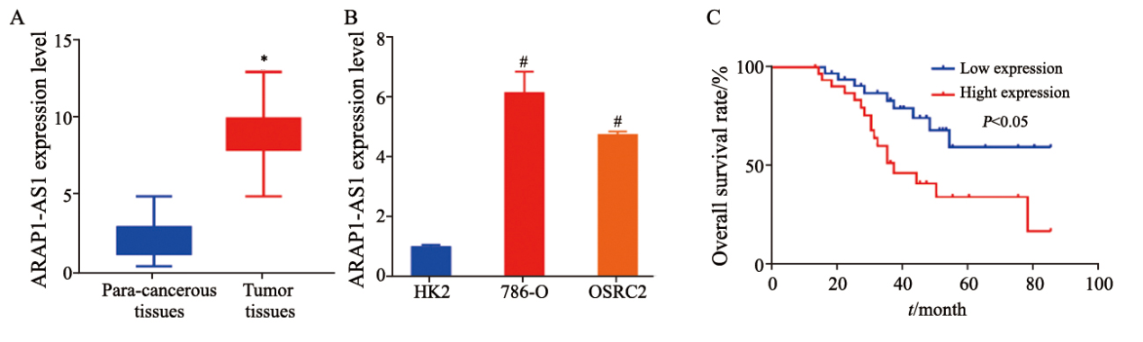
Fig. 2
ARAP1-AS1 expression in ccRCC tissues and cell lines and survival analysis of the patients
A: ccRCC tissues; B: ccRCC cell lines; C: Survival analysis; *: P<0.05, compared with para-cancerous tissues; #: P<0.05, compared with HK2.
Other Images/Table from this Article
|