| [1] |
FILHO A M, LAVERSANNE M, FERLAY J, et al. The GLOBOCAN 2022 cancer estimates: data sources, methods, and a snapshot of the cancer burden worldwide[J]. Int J Cancer, 2025, 156(7):1336-1346.
|
| [2] |
GOTO M, RYOO I, NAFFOUJE S, et al. Image-guided surgery with a new tumour-targeting probe improves the identification of positive margins[J]. EBioMedicine, 2022, 76: 103850.
|
| [3] |
ROSSOU C, ALAMPRITIS G, PATEL B. Reducing re-excision rates in breast conserving surgery with Margin Probe: systematic review[J]. Br J Surg, 2024, 111(1): znad335.
|
| [4] |
REID V J, FALK J S, POLICE A M, et al. Minimizing re-excision after breast conserving surgery-a review of radiofrequency spectroscopy for real-time, intraoperative margin assessment[J]. Expert Rev Med Devices, 2021, 18(11): 1057-1068.
|
| [5] |
VERONESI U, CASCINELLI N, MARIANI L, et al. Twenty-year follow-up of a randomized study comparing breast-conserving surgery with radical mastectomy for early breast cancer[J]. N Engl J Med, 2002, 347(16): 1227-1232.
|
| [6] |
HOFFMAN A, ASHKENAZI I. The efficiency of MarginProbe in detecting positive resection margins in epithelial breast cancer following breast conserving surgery[J]. Eur J Surg Oncol, 2022, 48(7): 1498-1502.
doi: 10.1016/j.ejso.2022.02.021
pmid: 35219544
|
| [7] |
MCCAHILL L E, SINGLE R M, AIELLO BOWLES E J, et al. Variability in reexcision following breast conservation surgery[J]. JAMA, 2012, 307(5): 467-475.
doi: 10.1001/jama.2012.43
pmid: 22298678
|
| [8] |
MORROW M, HARRIS J R, SCHNITT S J. Surgical margins in lumpectomy for breast cancer: bigger is not better[J]. N Engl J Med, 2012, 367(1): 79-82.
|
| [9] |
ST JOHN E R, AL-KHUDAIRI R, ASHRAFIAN H, et al. Diagnostic accuracy of intraoperative techniques for margin assessment in breast cancer surgery: a meta-analysis[J]. Ann Surg, 2017, 265(2): 300-310.
doi: 10.1097/SLA.0000000000001897
pmid: 27429028
|
| [10] |
RACZ J M, GLASGOW A E, KEENEY G L, et al. Intraoperative pathologic margin analysis and re-excision to minimize reoperation for patients undergoing breast-conserving surgery[J]. Ann Surg Oncol, 2020, 27(13): 5303-5311.
|
| [11] |
TANGSIRAPAT V, KENGSAKUL M, UDOMKARNJANANUN S, et al. Surgical margin status outcome of intraoperative indocyanine green fluorescence-guided laparoscopic hepatectomy in liver malignancy: a systematic review and meta-analysis[J]. BMC Surg, 2024, 24(1): 181.
doi: 10.1186/s12893-024-02469-1
pmid: 38867212
|
| [12] |
HISADA T, SAWAKI M, ISHIGURO J, et al. Impact of intraoperative specimen mammography on margins in breast-conserving surgery[J]. Mol Clin Oncol, 2016, 5(3): 269-272.
doi: 10.3892/mco.2016.948
pmid: 27588192
|
| [13] |
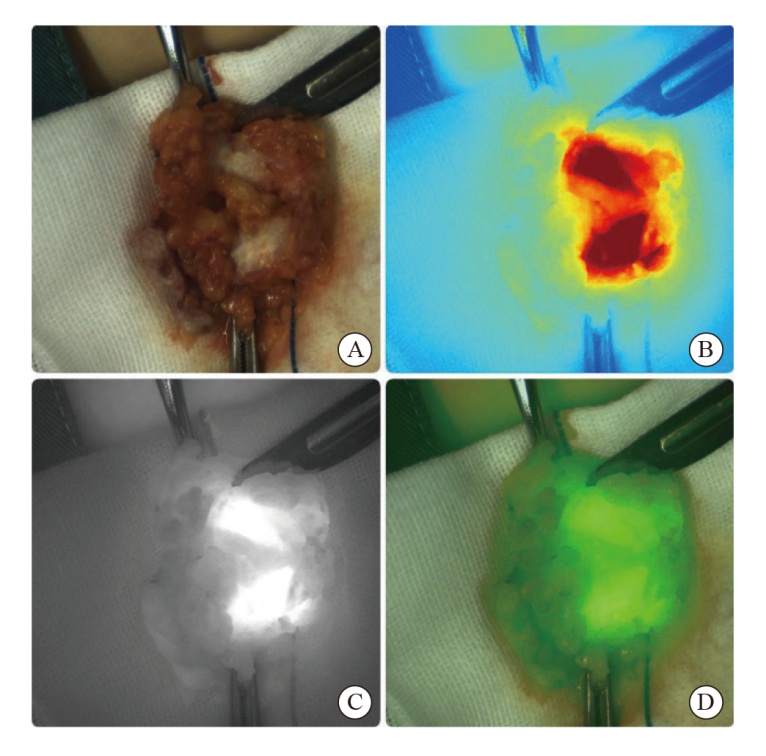
POP F C, VEYS I, VANKERCKHOVE S, et al. Absence of residual fluorescence in the surgical bed at near-infrared fluorescence imaging predicts negative margins at final pathology in patients treated with breast-conserving surgery for breast cancer[J]. Eur J Surg Oncol, 2021, 47(2): 269-275.
|
| [14] |
KEDRZYCKI M S, LEILOGLOU M, CHALAU V, et al. The impact of temporal variation in indocyanine green administration on tumor identification during fluorescence guided breast surgery[J]. Ann Surg Oncol, 2021, 28(10): 5617-5625.
doi: 10.1245/s10434-021-10503-2
pmid: 34347221
|
| [15] |
WANG Y B, JIAO W, YIN Z C, et al. Application of near-infrared fluorescence imaging in the accurate assessment of surgical margins during breast-conserving surgery[J]. World J Surg Oncol, 2022, 20(1): 357.
doi: 10.1186/s12957-022-02827-4
pmid: 36352391
|
| [16] |
BOICHENKO E, KIRSANOV D. Optical spectroscopy and chemometrics in intraoperative tumor margin assessment[J]. Trac Trends Anal Chem, 2023, 160: 116955.
|
 ), WANG Guangqing1, ZHENG Yan2, TANG Qin1, CHEN Fei1, YU Xudong1, XU Shengqi1, TANG Fayang3, ZHU Jibiao1(