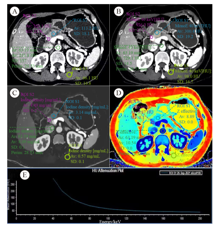
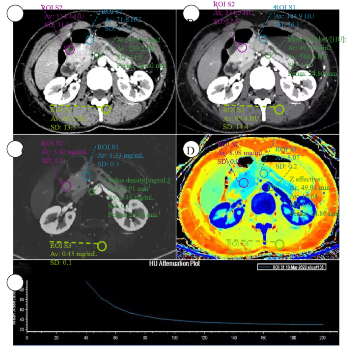
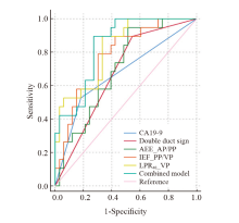
| [1] |
WHITCOMB D C, FRULLONI L, GARG P, et al. Chronic pancreatitis: an international draft consensus proposal for a new mechanistic definition[J]. Pancreatology, 2016, 16(2): 218-224.
doi: 10.1016/j.pan.2016.02.001
pmid: 26924663
|
| [2] |
SINGH V K, YADAV D, GARG P K. Diagnosis and management of chronic pancreatitis: a review[J]. JAMA, 2019, 322(24): 2422-2434.
doi: 10.1001/jama.2019.19411
pmid: 31860051
|
| [3] |
LIAO Q, ZHAO Y P, WU W W, et al. Diagnosis and treatment of chronic pancreatitis[J]. Hepatobiliary Pancreat Dis Int, 2003, 2(3): 445-448.
|
| [4] |
KIM T, MURAKAMI T, TAKAMURA M, et al. Pancreatic mass due to chronic pancreatitis: correlation of CT and MR imaging features with pathologic findings[J]. AJR Am J Roentgenol, 2001, 177(2): 367-371.
doi: 10.2214/ajr.177.2.1770367
|
| [5] |
KIRKEGÅRD J, MORTENSEN F V, CRONIN-FENTON D. Chronic pancreatitis and pancreatic cancer risk: a systematic review and meta-analysis[J]. Am J Gastroenterol, 2017, 112(9): 1366-1372.
doi: 10.1038/ajg.2017.218
pmid: 28762376
|
| [6] |
LUETMER P H, STEPHENS D H, WARD E M. Chronic pancreatitis: reassessment with current CT[J]. Radiology, 1989, 171(2): 353-357.
doi: 10.1148/radiology.171.2.2704799
pmid: 2704799
|
| [7] |
WOLSKE K M, PONNATAPURA J, KOLOKYTHAS O, et al. Chronic pancreatitis or pancreatic tumor? A problem-solving approach[J]. Radiographics, 2019, 39(7): 1965-1982.
doi: 10.1148/rg.2019190011
pmid: 31584860
|
| [8] |
GANDHI S, DE LA FUENTE J, MURAD M H, et al. Chronic pancreatitis is a risk factor for pancreatic cancer, and incidence increases with duration of disease: a systematic review and meta-analysis[J]. Clin Transl Gastroenterol, 2022, 13(3): e00463.
doi: 10.14309/ctg.0000000000000463
|
| [9] |
国家卫生健康委办公厅. 胰腺癌诊疗指南(2022年版)[J]. 临床肝胆病杂志, 2022, 38(5): 1006-1030.
|
|
General Office of National Health Commission. Standard for diagnosis and treatment of pancreatic cancer (2022 edition)[J]. J Clin Hepatol, 2022, 38(5): 1006-1030.
|
| [10] |
ASLAN S, NURAL M S, CAMLIDAG I, et al. Efficacy of perfusion CT in differentiating of pancreatic ductal adenocarcinoma from mass-forming chronic pancreatitis and characterization of isoattenuating pancreatic lesions[J]. Abdom Radiol, 2019, 44(2): 593-603.
doi: 10.1007/s00261-018-1776-9
|
| [11] |
SANDRASEGARAN K, NUTAKKI K, TAHIR B, et al. Use of diffusion-weighted MRI to differentiate chronic pancreatitis from pancreatic cancer[J]. AJR Am J Roentgenol, 2013, 201(5): 1002-1008.
doi: 10.2214/AJR.12.10170
|
| [12] |
ELSHERIF S B, VIRARKAR M, JAVADI S, et al. Pancreatitis and PDAC: association and differentiation[J]. Abdom Radiol (NY), 2020, 45(5): 1324-1337.
doi: 10.1007/s00261-019-02292-w
pmid: 31705251
|
| [13] |
SEICEAN A, BADEA R, MOLDOVAN-POP A, et al. Harmonic contrast-enhanced endoscopic ultrasonography forthe guidance of fine-needle aspiration in solid pancreatic masses[J]. Ultraschall Med, 2017, 38(2): 174-182.
doi: 10.1055/s-0035-1553496
|
| [14] |
FACCIORUSSO A, MARTINA M, BUCCINO R V, et al. Diagnostic accuracy of fine-needle aspiration of solid pancreatic lesions guided by endoscopic ultrasound elastography[J]. Ann Gastroenterol, 2018, 31(4): 513-518.
|
| [15] |
FRITSCHER-RAVENS A, BRAND L, KNÖFEL W T, et al. Comparison of endoscopic ultrasound-guided fine needle aspiration for focal pancreatic lesions in patients with normal parenchyma and chronic pancreatitis[J]. Am J Gastroenterol, 2002, 97(11): 2768-2775.
doi: 10.1111/j.1572-0241.2002.07020.x
|
| [16] |
BEER L, TOEPKER M, BA-SSALAMAH A, et al. Objective and subjective comparison of virtual monoenergetic vs polychromatic images in patients with pancreatic ductal adenocarcinoma[J]. Eur Radiol, 2019, 29(7): 3617-3625.
doi: 10.1007/s00330-019-06116-9
|
| [17] |
SHI H Y, LU Z P, LI M N, et al. Dual-energy CT iodine concentration to evaluate postoperative pancreatic fistula after pancreatoduodenectomy[J]. Radiology, 2022, 304(1): 65-72.
doi: 10.1148/radiol.212173
|
| [18] |
YIN Q H, ZOU X N, ZAI X D, et al. Pancreatic ductal adenocarcinoma and chronic mass-forming pancreatitis: differentiation with dual-energy MDCT in spectral imaging mode[J]. Eur J Radiol, 2015, 84(12): 2470-2476.
doi: 10.1016/j.ejrad.2015.09.023
pmid: 26481480
|
| [19] |
GROßE HOKAMP N, MAINTZ D, SHAPIRA N, et al. Technical background of a novel detector-based approach to dual-energy computed tomography[J]. Diagn Interv Radiol, 2020, 26(1): 68-71.
doi: 10.5152/dir.2019.19136
pmid: 31904573
|
| [20] |
ZHANG H, MENG Y H, LI Q, et al. Two nomograms for differentiating mass-forming chronic pancreatitis from pancreatic ductal adenocarcinoma in patients with chronic pancreatitis[J]. Eur Radiol, 2022, 32(9): 6336-6347.
doi: 10.1007/s00330-022-08698-3
pmid: 35394185
|
| [21] |
YEH B M, SHEPHERD J A, WANG Z J, et al. Dual-energy and low-kVp CT in the abdomen[J]. AJR Am J Roentgenol, 2009, 193(1): 47-54.
doi: 10.2214/AJR.09.2592
|
| [22] |
GHASEMI SHAYAN R, OLADGHAFFARI M, SAJJADIAN F, et al. Image quality and dose comparison of single-energy CT (SECT) and dual-energy CT (DECT)[J]. Radiol Res Pract, 2020, 2020: 1403957.
|
| [23] |
SAKABE D, FUNAMA Y, TAGUCHI K, et al. Image quality characteristics for virtual monoenergetic images using dual-layer spectral detector CT: comparison with conventional tube-voltage images[J]. Phys Med, 2018, 49: 5-10.
doi: S1120-1797(18)30447-2
pmid: 29866343
|
| [24] |
REN S, ZHANG J J, CHEN J Y, et al. Evaluation of texture analysis for the differential diagnosis of mass-forming pancreatitis from pancreatic ductal adenocarcinoma on contrast-enhanced CT images[J]. Front Oncol, 2019, 9: 1171.
doi: 10.3389/fonc.2019.01171
pmid: 31750254
|
| [25] |
SU S B, QIN S Y, CHEN W, et al. Carbohydrate antigen 19-9 for differential diagnosis of pancreatic carcinoma and chronic pancreatitis[J]. World J Gastroenterol, 2015, 21(14): 4323-4333.
doi: 10.3748/wjg.v21.i14.4323
|
| [26] |
SINGH S, TANG S J, SREENARASIMHAIAH J, et al. The clinical utility and limitations of serum carbohydrate antigen (CA19-9) as a diagnostic tool for pancreatic cancer and cholangiocarcinoma[J]. Dig Dis Sci, 2011, 56(8): 2491-2496.
doi: 10.1007/s10620-011-1709-8
|
 ), XIE Tiansong1, CHEN Lei2, ZHANG Zehua2, ZHOU Zhengrong1,2(