Welcome to China Oncology,
China Oncology ›› 2024, Vol. 34 ›› Issue (9): 838-847.doi: 10.19401/j.cnki.1007-3639.2024.09.004
• Article • Previous Articles Next Articles
CHEN Yijun( ), LIU Yuhang, DUAN Haibo, WANG Xiongjun()
Received:2024-04-20
Revised:2024-09-09
Online:2024-09-30
Published:2024-10-11
Contact:
WANG Xiongjun
Share article
CLC Number:
CHEN Yijun, LIU Yuhang, DUAN Haibo, WANG Xiongjun. Functional and mechanistic of AGPAT5 in liver cancer[J]. China Oncology, 2024, 34(9): 838-847.
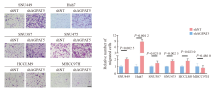
Fig. 1
Knockdown of AGPAT5 promotes HCC cells migration Transwell migration assays were assessed in AGPAT5-knockdown HCC cell lines. Representative images (scale bar: 100 μm) and statistical analyses of the migrated cells are shown."
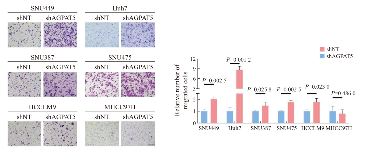

Fig. 2
Depletion of AGPAT5 induces tolerance to serum-free starvation in HCC cells A, B: SNU449 and Huh7 cells stably expressing shNT or shAGPAT5 were treated with or without FBS starvation in the indicated times, and then, cell number was assessed. C: Colony formation assays were performed in AGPAT5-depleted SNU449 and Huh7 cells. Representative images and statistical analyses of the colony cells are shown."
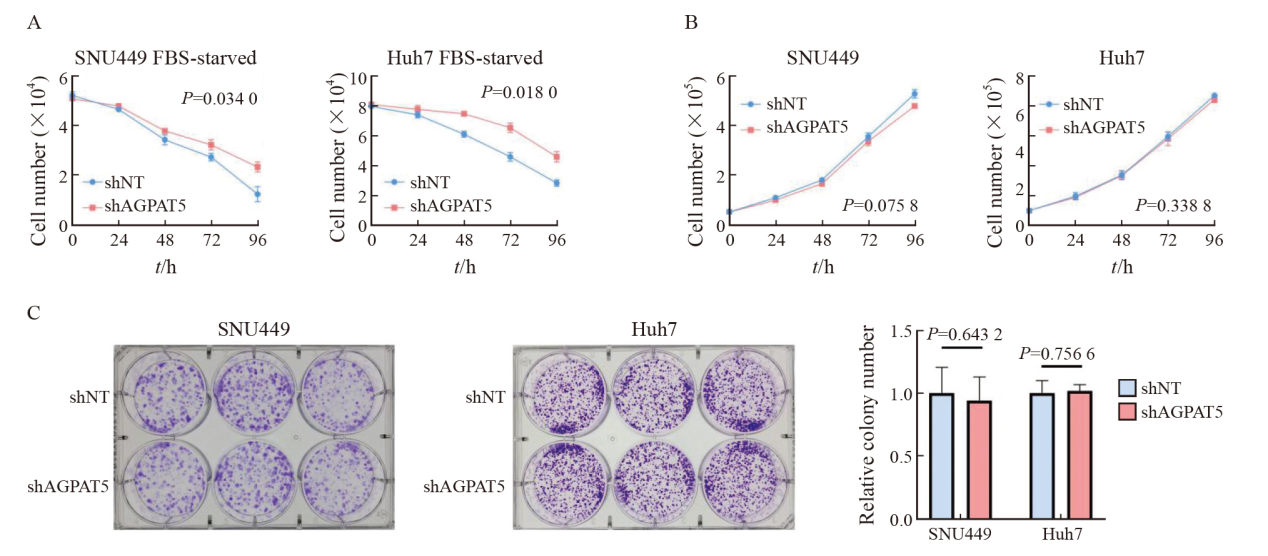
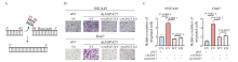
Fig. 3
AGPAT5 inactivation does not affect the migration of HCC cells A: Construct the enzyme deficiency plasmid. B, C: Transwell migration assays were performed in AGPAT5-depleted SNU449 and Huh7 cells rescued with EV or rAGPAT5-WT or rAGPAT5-ED. Representative images (scale bar: 100 μm) and statistical analyses of the migrated cells are shown."
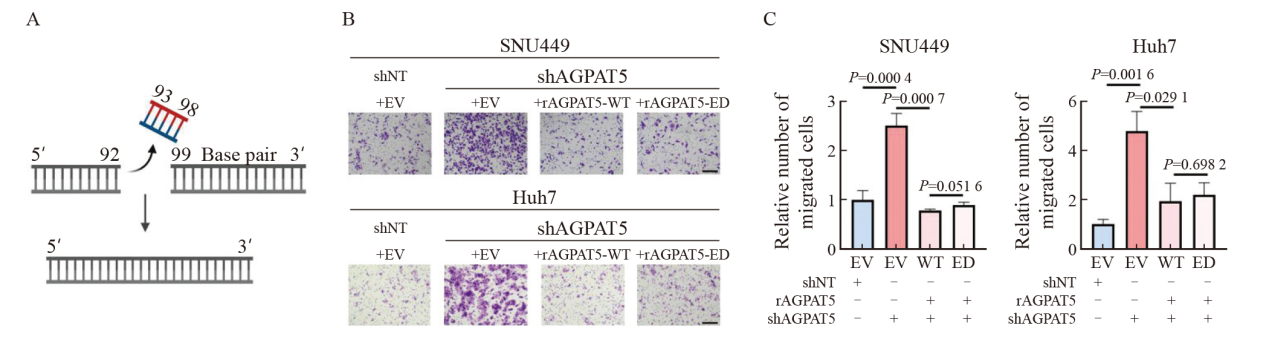

Fig. 4
Depletion of AGPAT5 promotes the metastasis of HCC cells in mice A, B, C: SNU449-shNT cells or SNU449-shAGPAT5 cells were implanted into randomized athymic nude mice via a tail vein injection (6 mice per group). Then, after 45 d of inoculation, the representative images of these implanted mice was carried out, the body weight of mice was quantified. The representative images of lung and liver metastasis are presented. D, E, F: Representative images of liver and lung tissues dissected 45 d after the inoculation and H-E and Ki-67 stained metastatic nodules are presented (scale bar: 50 μm) and statistical analyses of metastatic nodules are shown."
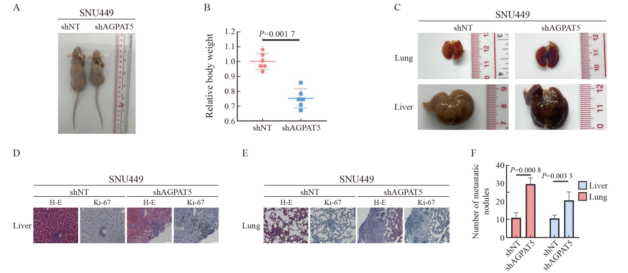

Fig. 5
AGPAT5 may inhibit the migration of HCC cells by suppressing FBL A: Mass spectrometry analyses of AGPAT5-associated proteins were performed in SNU449 cells stably expressing FLAG-AGPAT5. The precursor ion was fragmented by collision-induced dissociation and analyzed in an ion trap. The database search engine (Andromeda) score was matched to the identified peptide. B: SNU449 and Huh7 cells were treated with or without FBS for 1 h. C, D: Transwell migration assays were performed in AGPAT5-depleted or AGPAT5-overexpressed or FBL-depleted SNU449 and Huh7 cells. Representative images (scale bar: 100 μm) and statistical analyses of the migrated cells are shown. E, F: Transwell assays revealed that FBL depletion abrogated the promoted migration caused by AGPAT5 knockdown in SNU449 and Huh7 cells. G, H: HCC cell lysate to test the relative expression level of AGPAT5 and FBL. IB: immunoblot; IP: immunoprecipitation."
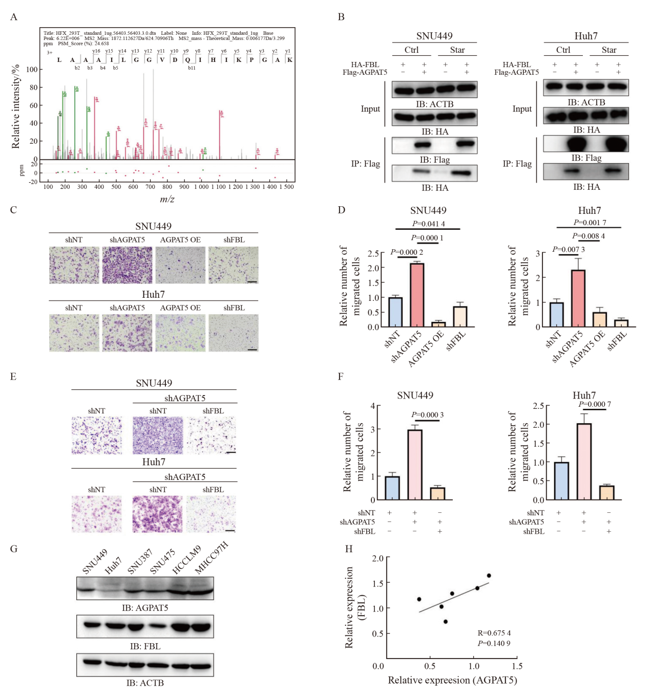

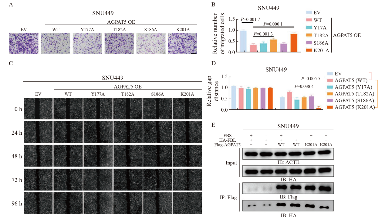
Fig. 6
K201 of AGPAT5 is the key site for FBL repression A, B, C, D: Transwell migration assays and wound healing assays of SNU449-AGPAT5 (WT), SNU449-AGPAT5 (Y177A), SNU449-AGPAT5 (T182A), SNU449-AGPAT5 (S186A) and SNU449-AGPAT5 (K201A) cells. Representative images (A) (scale bar: 100 μm) and statistical analyses of the migrated cells are shown. Representative images (C) (scale bar: 2.5 mm) and statistical analyses of gap distance are shown. E: SNU449 cells were treated with or without FBS for 1 h."
| [1] | SUNG H, FERLAY J, SIEGEL R L, et al. Global cancer statistics 2020: GLOBOCAN estimates of incidence and mortality worldwide for 36 cancers in 185 countries[J]. CA Cancer J Clin, 2021, 71(3): 209-249. |
| [2] | BRAY F, FERLAY J, SOERJOMATARAM I, et al. Global cancer statistics 2018: GLOBOCAN estimates of incidence and mortality worldwide for 36 cancers in 185 countries[J]. CA Cancer J Clin, 2018, 68(6): 394-424. |
| [3] | BUDNY A, KOZŁOWSKI P, KAMIŃSKA M, et al. Epidemiology and risk factors of hepatocellular carcinoma[J]. Pol Merkur Lekarski, 2017, 43(255): 133-139. |
| [4] |
FORNER A, LLOVET J M, BRUIX J. Hepatocellular carcinoma[J]. Lancet, 2012, 379(9822): 1245-1255.
doi: 10.1016/S0140-6736(11)61347-0 pmid: 22353262 |
| [5] |
TSILIMIGRAS D I, BRODT P, CLAVIEN P A, et al. Liver metastases[J]. Nat Rev Dis Primers, 2021, 7(1): 27.
doi: 10.1038/s41572-021-00261-6 pmid: 33859205 |
| [6] |
LEE Y T, GEER D A. Primary liver cancer: pattern of metastasis[J]. J Surg Oncol, 1987, 36(1): 26-31.
pmid: 3041113 |
| [7] | VALENTINE W J, YANAGIDA K, KAWANA H, et al. Update and nomenclature proposal for mammalian lysophospholipid acyltransferases, which create membrane phospholipid diversity[J]. J Biol Chem, 2022, 298(1): 101470. |
| [8] | KARAGIOTA A, CHACHAMI G, PARASKEVA E. Lipid metabolism in cancer: the role of acylglycerolphosphate acyltransferases (AGPATs)[J]. Cancers, 2022, 14(1): 228. |
| [9] |
PRASAD S S, GARG A, AGARWAL A K. Enzymatic activities of the human AGPAT isoform 3 and isoform 5: localization of AGPAT5 to mitochondria[J]. J Lipid Res, 2011, 52(3): 451-462.
doi: 10.1194/jlr.M007575 pmid: 21173190 |
| [10] | VARGAS T, MORENO-RUBIO J, HERRANZ J, et al. ColoLipidGene: signature of lipid metabolism-related genes to predict prognosis in stage-Ⅱ colon cancer patients[J]. Oncotarget, 2015, 6(9): 7348-7363. |
| [11] | NIESPOREK S, DENKERT C, WEICHERT W, et al. Expression of lysophosphatidic acid acyltransferase beta (LPAAT-beta) in ovarian carcinoma: correlation with tumour grading and prognosis[J]. Br J Cancer, 2005, 92(9): 1729-1736. |
| [12] | BLASKOVICH M A, YENDLURI V, LAWRENCE H R, et al. Lysophosphatidic acid acyltransferase beta regulates mTOR signaling[J]. PLoS One, 2013, 8(10): e78632. |
| [13] |
DÓRIA M L, RIBEIRO A S, WANG J, et al. Fatty acid and phospholipid biosynthetic pathways are regulated throughout mammary epithelial cell differentiation and correlate to breast cancer survival[J]. FASEB J, 2014, 28(10): 4247-4264.
doi: 10.1096/fj.14-249672 pmid: 24970396 |
| [14] | ZHANG D P, SHI R C, XIANG W, et al. The Agpat4/LPA axis in colorectal cancer cells regulates antitumor responses via p38/p65 signaling in macrophages[J]. Signal Transduct Target Ther, 2020, 5(1): 24. |
| [15] | SUMANTRAN V N, MISHRA P, SUDHAKAR N. Microarray analysis of differentially expressed genes regulating lipid metabolism during melanoma progression[J]. Indian J Biochem Biophys, 2015, 52(2): 125-131. |
| [16] | LI M, ZHAO Z W, ZHANG Y, et al. Over-expression of Ephb4 is associated with carcinogenesis of gastric cancer[J]. Dig Dis Sci, 2011, 56(3): 698-706. |
| [17] | SONG L, YANG J, DUAN P, et al. MicroRNA-24 inhibits osteosarcoma cell proliferation both in vitro and in vivo by targeting LPAATβ[J]. Arch Biochem Biophys, 2013, 535(2): 128-135. |
| [18] |
YANG J F, XIANG C P, LIU J M. Clinical significance of combining salivary mRNAs and carcinoembryonic antigen for ovarian cancer detection[J]. Scand J Clin Lab Invest, 2021, 81(1): 39-45.
doi: 10.1080/00365513.2020.1852478 pmid: 33300816 |
| [19] | ZANG J, SUN J J, XIU W C, et al. Low expression of AGPAT5 is associated with clinical stage and poor prognosis in colorectal cancer and contributes to tumour progression[J]. Clin Med Insights Oncol, 2022, 16: 11795549221137399. |
| [20] | YING Q, ANSONG E, DIAMOND A M, et al. Abstract 1197: Selenium-binding protein 1-mediated tumor suppression is associated with alterations of lipid/glucose metabolic pathways in vivo[J]. Cancer Res, 2015, 75(15_Supplement): 1197. |
| [21] | WEN P Z, WANG R, XING Y Q, et al. The prognostic value of the GPAT/AGPAT gene family in hepatocellular carcinoma and its role in the tumor immune microenvironment[J]. Front Immunol, 2023, 14: 1026669. |
| [22] | FAUBERT B, SOLMONSON A, DEBERARDINIS R J. Metabolic reprogramming and cancer progression[J]. Science, 2020, 368(6487): eaaw5473. |
| [23] | MARTIN-PEREZ M, URDIROZ-URRICELQUI U, BIGAS C, et al. The role of lipids in cancer progression and metastasis[J]. Cell Metab, 2022, 34(11): 1675-1699. |
| [24] |
ZHANG Q, YAO D Q, RAO B, et al. The structural basis for the phospholipid remodeling by lysophosphatidylcholine acyltransferase 3[J]. Nat Commun, 2021, 12(1): 6869.
doi: 10.1038/s41467-021-27244-1 pmid: 34824256 |
| [25] | ZHU H W, YU H, ZHOU H, et al. Elevated nuclear PHGDH synergistically functions with cMyc to reshape the immune microenvironment of liver cancer[J]. Adv Sci, 2023, 10(17): e2205818. |
| [26] |
YU H, SHI T Z, YAO L L, et al. Elevated nuclear PIGL disrupts the cMyc/BRD4 axis and improves PD-1 blockade therapy by dampening tumor immune evasion[J]. Cell Mol Immunol, 2023, 20(8): 867-880.
doi: 10.1038/s41423-023-01048-3 pmid: 37280393 |
| [27] |
MARCEL V, GHAYAD S E, BELIN S, et al. p53 acts as a safeguard of translational control by regulating fibrillarin and rRNA methylation in cancer[J]. Cancer Cell, 2013, 24(3): 318-330.
doi: 10.1016/j.ccr.2013.08.013 pmid: 24029231 |
| [28] | SUN X R, GAO C W, XU X, et al. FBL promotes cancer cell resistance to DNA damage and BRCA1 transcription via YBX1[J]. EMBO Rep, 2023, 24(9): e56230. |
| [29] | LIU Y Z, SHI Q L, LIU Y F, et al. Fibrillarin reprograms glucose metabolism by driving the enhancer-mediated transcription of PFKFB4 in liver cancer[J]. Cancer Lett, 2024, 602: 217190. |
| [30] |
ZHANG J, YANG G, LI Q, et al. Increased fibrillarin expression is associated with tumor progression and an unfavorable prognosis in hepatocellular carcinoma[J]. Oncol Lett, 2021, 21(2): 92.
doi: 10.3892/ol.2020.12353 pmid: 33376525 |
| [1] | WEN Ziqiang, LAN Junliang, ZHOU Bo, XU Qiwei. PARP1 promotes the progression of hepatocellular carcinoma by regulating expression of POU2F2 [J]. China Oncology, 2024, 34(9): 848-856. |
| [2] | XIAO Feng, XU Tonglin, ZHU Lin, XIAO Jingwen, WU Tianqi, GU Chunyan. Significance of infiltration of M1 tumor-associated macrophages in hepatocellular carcinoma [J]. China Oncology, 2024, 34(8): 726-733. |
| [3] | ZHAO Haichao, GAO Qiang. Progress in research, diagnosis, and treatment of hepatocellular carcinoma in 2022 [J]. China Oncology, 2023, 33(4): 315-326. |
| [4] | HU Keshu, LIU Wenfeng, ZHANG Feng, QUAN Bing, YIN Xin. Mechanism of LINC00601 in regulating sensitivity of hepatocellular carcinoma cells to oxaliplatin chemotherapy [J]. China Oncology, 2023, 33(1): 25-35. |
| [5] | LIU Xueping, MU Nai, LI Xiaochun, WANG Ruisi, TAN Bangxian. Gingival metastasis from primary hepatocellular carcinoma: a case report [J]. China Oncology, 2023, 33(1): 78-80. |
| [6] | CHENG Yicai, DU Zhenhua, FAN Zhijuan, FENG Lan, CAO Pengbo, ZHOU Gangqiao. Plasma exosomal CD48 protein as a candidate diagnostic biomarker for hepatocellular carcinoma [J]. China Oncology, 2022, 32(11): 1074-1083. |
| [7] | GUO Tao, SONG Ning, SUN Yuqi, TIAN Jiaqi, TANG Jie, SUN Rongqi, JIANG Yingying. Effect of long non-coding RNA LINC00671 on biological behavior of hepatocellular carcinoma and its mechanism [J]. China Oncology, 2022, 32(10): 990-999. |
| [8] | YU Hang, LIU Wensheng, ZHANG Ning, LIU Haikuan, CHEN Luohai, YAO Wang, FAN Wenzhe, LI Jiaping, CHEN Jie, WANG Yu. The efficacy and safety analysis of transarterial embolization in the treatment of cystic neuroendocrine neoplasm liver metastasis [J]. China Oncology, 2022, 32(9): 794-799. |
| Viewed | ||||||
|
Full text |
|
|||||
|
Abstract |
|
|||||
沪ICP备12009617
Powered by Beijing Magtech Co. Ltd