Welcome to China Oncology,
China Oncology ›› 2023, Vol. 33 ›› Issue (1): 36-44.doi: 10.19401/j.cnki.1007-3639.2023.01.004
• Article • Previous Articles Next Articles
ZOU Chunyuan( ), XU Xiaofeng, LU Renquan(), GUO Lin
Received:2022-09-28
Revised:2022-11-02
Online:2023-01-30
Published:2023-02-13
Contact:
LU Renquan
Share article
CLC Number:
ZOU Chunyuan, XU Xiaofeng, LU Renquan, GUO Lin. Detection of p53, PGP9.5, SOX2, GAGE7, GBU4-5 and MAGE A1 protein levels in lung cancer tissues and peripheral blood and their clinical value[J]. China Oncology, 2023, 33(1): 36-44.
Tab. 1
Medical cut-off values of 6 indicators"
| Autoantibody | P53 | PGP9.5 | SOX2 | GAGE7 | GBU4-5 | MAGE A1 |
|---|---|---|---|---|---|---|
| Cut-off value/(U·mL-1) | 13.1 | 11.1 | 10.3 | 14.4 | 7.0 | 11.9 |
Tab. 2
Comparison of positive protein expression in tumor tissues and adjacent tissues and serum [n (%)]"
| Item | p53 | PGP9.5 | SOX2 | GAGE7 | GBU4-5 | MAGE A1 |
|---|---|---|---|---|---|---|
| Tumor tissue | 41 (41.0) | 45 (45.0) | 32 (32.0) | 36 (36.0) | 37 (37.0) | 39 (39.0) |
| Adjacent tissue | 3 (3.0) | 4 (4.0) | 2 (2.0) | 1 (1.0) | 1 (1.0) | 1 (1.0) |
| Serum | 35 (35.0) | 46 (46.0) | 30 (30.0) | 34 (34.0) | 35 (35.0) | 40 (40.0) |
| χ2 value | 44.477 | 31.558 | 23.926 | 29.587 | 30.429 | 33.325 |
| P value | <0.001 | <0.001 | <0.001 | <0.001 | <0.001 | <0.001 |
Tab. 3
Comparison of protein relative expression levels in tumor tissues and adjacent tissues and serum expression levels ($\bar{x}±s$)"
| Item | p53 | PGP9.5 | SOX2 | GAGE7 | GBU4-5 | MAGE A1 |
|---|---|---|---|---|---|---|
| Tumor tissue | 0.52±0.13 | 0.48±0.14 | 0.39±0.11 | 0.35±0.12 | 0.29±0.08 | 0.42±0.19 |
| Adjacent tissue | 0.16±0.05 | 0.15±0.04 | 0.09±0.02 | 0.10±0.03 | 0.15±0.06 | 0.18±0.07 |
| Serum | 6.47±11.81 | 1.16±3.75 | 5.61±9.14 | 3.36±5.52 | 1.81±2.84 | 2.31±4.82 |
| F value | 5.038 | 8.137 | 5.121 | 4.389 | 5.009 | 5.001 |
| P value | <0.001 | <0.001 | <0.001 | <0.001 | <0.001 | <0.001 |
Tab. 4
Relationship between positive and relative expression levels of protein in tumor tissues and clinicopathological characteristics of tumors [Quantification (n)]"
| Item | Case n | p53 | PGP9.5 | SOX2 | GAGE7 | GBU4-5 | MAGE A1 |
|---|---|---|---|---|---|---|---|
| Gender | |||||||
| Men | 60 | 0.53±0.14 (25) | 0.46±0.12 (27) | 0.38±0.12 (19) | 0.33±0.09 (24) | 0.28±0.11 (25) | 0.41±0.16 (26) |
| Women | 40 | 0.51±0.13 (16) | 0.49±0.17 (18) | 0.39±0.16 (13) | 0.36±0.12 (12) | 0.29±0.12 (12) | 0.43±0.15 (13) |
| t/χ2 value | 0.122 (0.028) | 0.163 (0.000) | 0.155 (0.293) | 0.096 (1.042) | 0.121 (1.401) | 0.113 (1.184) | |
| P value | 0.869 (0.868) | 0.789 (1.000) | 0.825 (0.588) | 0.968 (0.307) | 0.923 (0.236) | 0.869 (0.277) | |
| Age/year | |||||||
| <60.5 | 42 | 0.52±0.14 (17) | 0.47±0.16 (20) | 0.37±0.12 (13) | 0.34±0.10 (13) | 0.27±0.09 (13) | 0.40±0.18 (14) |
| ≥60.5 | 58 | 0.54±0.16 (24) | 0.49±0.19 (25) | 0.40±0.16 (19) | 0.36±0.12 (23) | 0.30±0.10 (24) | 0.43±0.18 (25) |
| t/χ2 value | 0.203 (0.008) | 0.185 (0.201) | 0.252 (0.037) | 0.156 (0.801) | 0.232 (1.136) | 0.257 (0.977) | |
| P value | 0.724 (0.928) | 0.769 (0.654) | 0.689 (0.848) | 0.868 (0.371) | 0.822 (0.286) | 0.786 (0.323) | |
| Tumor diameter/cm | |||||||
| <3.5 | 46 | 0.51±0.13 (18) | 0.47±0.16 (19) | 0.38±0.12 (14) | 0.35±0.11 (14) | 0.29±0.11 (14) | 0.42±0.16 (15) |
| ≥3.5 | 54 | 0.55±0.19 (23) | 0.49±0.18 (26) | 0.40±0.16 (18) | 0.36±0.13 (22) | 0.31±0.16 (23) | 0.43±0.18 (24) |
| t/χ2 value | 0.320 (0.123) | 0.264 (0.470) | 0.358 (0.096) | 0.265 (1.145) | 0.321 (1.575) | 0.367 (1.463) | |
| P value | 0.764 (0.726) | 0.806 (0.493) | 0.702 (0.757) | 0.802 (0.285) | 0.763 (0.209) | 0.712 (0.227) | |
| Pathologic type | |||||||
| SCLC | 20 | 0.52±0.15 (8) | 0.46±0.18 (11) | 0.37±0.12 (6) | 0.33±0.08 (8) | 0.28±0.12 (8) | 0.41±0.19 (9) |
| NSCLC | 80 | 0.53±0.16 (33) | 0.49±0.18 (34) | 0.41±0.16 (26) | 0.36±0.12 (28) | 0.30±0.13 (29) | 0.43±0.21 (30) |
| t/χ2 value | 0.524 (0.010) | 0.326 (1.010) | 0.421 (0.046) | 0.326 (0.174) | 0.402 (0.097) | 0.456 (0.378) | |
| P value | 0.523 (0.919) | 0.659 (0.315) | 0.603 (0.830) | 0.721 (0.677) | 0.695 (0.756) | 0.636 (0.539) | |
| TNM | |||||||
| Ⅰ | 25 | 0.48±0.15 (3) | 0.46±0.18 (4) | 0.36±0.19 (2) | 0.31±0.08 (2) | 0.28±0.12 (2) | 0.40±0.17 (2) |
| Ⅱ | 45 | 0.53±0.16 (15) | 0.47±0.16 (15) | 0.38±0.12 (10) | 0.36±0.11 (13) | 0.30±0.13 (13) | 0.43±0.16 (13) |
| Ⅲa | 30 | 0.59±0.17 (23) | 0.52±0.20 (26) | 0.42±0.16 (20) | 0.39±0.13 (21) | 0.34±0.15 (22) | 0.46±0.19 (24) |
| F/χ2 value | 5.236 (25.562) | 5.524 (32.013) | 5.123 (25.163) | 4.659 (24.547) | 5.003 (27.280) | 5.212 (32.231) | |
| P value | 0.000 (0.000) | 0.000 (0.000) | 0.000 (0.000) | 0.013 (0.000) | 0.007 (0.000) | 0.003 (0.000) | |
| Differentiation degree | |||||||
| Low | 40 | 0.60±0.18 (31) | 0.53±0.18 (34) | 0.43±0.18 (25) | 0.41±0.16 (26) | 0.33±0.16 (27) | 0.47±0.21 (29) |
| Middle | 35 | 0.55±0.17 (8) | 0.48±0.16 (8) | 0.38±0.16 (5) | 0.37±0.16 (8) | 0.29±0.15 (8) | 0.44±0.18 (8) |
| High | 25 | 0.46±0.14 (2) | 0.45±0.18 (3) | 0.35±0.16 (2) | 0.33±0.15 (2) | 0.26±0.12 (2) | 0.40±0.15 (2) |
| F/χ2 value | 5.659 (38.047) | 5.857 (43.792) | 5.903 (28.765) | 5.524 (25.732) | 5.968 (27.986) | 6.003 (32.802) | |
| P value | 0.000 (0.000) | 0.000 (0.000) | 0.000 (0.000) | 0.001 (0.000) | 0.000 (0.000) | 0.000 (0.000) |
Tab. 5
Relationship between the positive rate and level of p53, PGP9.5, SOX2, GAGE7, GBU4-5, MAGE A1 in serum and the clinicopathological characteristics of tumors [Quantification (n)]"
| Item | Case n | p53 | PGP9.5 | SOX2 | GAGE7 | GBU4-5 | MAGE A1 |
|---|---|---|---|---|---|---|---|
| Gender | |||||||
| Men | 60 | 6.23±10.84 (22) | 1.11±3.12 (25) | 5.58±9.14 (17) | 3.23±5.09 (24) | 1.78±2.71 (22) | 2.43±4.76 (24) |
| Women | 40 | 6.51±9.73 (13) | 1.09±3.17 (21) | 5.62±8.96 (13) | 3.16±5.12 (10) | 1.69±2.62 (13) | 2.26±4.55 (16) |
| t/χ2 value | 0.132 (0.183) | 0.031 (1.134) | 0.022 (0.499) | 0.067 (2.406) | 0.165 (0.183) | 0.178 (0.000) | |
| P value | 0.896 (0.669) | 0.975 (0.287) | 0.983 (0.480) | 0.947 (0.121) | 0.869 (0.669) | 0.859 (1.000) | |
| Age/year | |||||||
| <60.5 | 42 | 6.12±10.14 (14) | 1.07±3.16 (19) | 5.37±9.12 (11) | 3.34±5.11 (11) | 1.67±2.59 (11) | 2.40±4.68 (13) |
| ≥60.5 | 58 | 6.04±10.16 (21) | 1.19±3.19 (27) | 5.40±8.76 (19) | 3.26±5.07 (23) | 1.70±2.60 (24) | 2.23±4.56 (27) |
| t/χ2 value | 0.039 (0.088) | 0.186 (0.017) | 0.017 (0.500) | 0.078 (1.968) | 0.057 (2.470) | 0.182 (2.470) | |
| P value | 0.969 (0.766) | 0.953 (0.896) | 0.987 (0.479) | 0.938 (0.161) | 0.955 (0.116) | 0.856 (0.116) | |
| Tumor diameter/cm | |||||||
| <3.5 | 46 | 6.21±9.73 (15) | 1.15±3.13 (21) | 5.38±9.15 (11) | 3.29±5.16 (13) | 1.59±2.61 (13) | 2.42±4.46 (15) |
| ≥3.5 | 54 | 6.15±10.12 (20) | 1.17±2.98 (25) | 5.43±8.22 (19) | 3.24±5.09 (21) | 1.62±2.55 (22) | 2.33±4.38 (25) |
| t/χ2 value | 0.030 (0.214) | 0.033 (0.004) | 0.029 (1.503) | 0.049 (1.250) | 0.058 (1.701) | 0.102 (1.939) | |
| P value | 0.976 (0.644) | 0.974 (0.949) | 0.977 (0.220) | 0.961 (0.263) | 0.954 (0.192) | 0.919 (0.164) | |
| Pathologic type | |||||||
| SCLC | 20 | 6.12±9.65 (6) | 1.18±3.22 (10) | 5.33±9.12 (4) | 3.33±5.08 (7) | 1.48±2.52 (5) | 2.41±4.19 (11) |
| NSCLC | 80 | 6.03±10.11 (29) | 1.22±2.89 (36) | 5.41±9.24 (26) | 2.26±5.12 (27) | 1.60±2.63 (30) | 2.32±4.27 (29) |
| t/χ2 value | 0.036 (0.275) | 0.054 (0.161) | 0.035 (1.191) | 0.837 (0.011) | 0.184 (1.099) | 0.085 (2.344) | |
| P value | 0.971 (0.600) | 0.957 (0.688) | 0.972 (0.275) | 0.405 (0.916) | 0.854 (0.295) | 0.933 (0.126) | |
| TNM | |||||||
| Ⅰ | 25 | 5.48±7.15 (1) | 0.86±2.16 (5) | 5.06±9.15 (1) | 2.41±3.08 (1) | 1.18±1.82 (2) | 2.12±2.13 (3) |
| Ⅱ | 45 | 6.13±10.16 (13) | 1.17±3.11 (13) | 5.28±8.92 (11) | 3.16±4.11 (11) | 1.35±2.13 (11) | 2.43±2.06 (11) |
| Ⅲa | 30 | 6.29±10.08 (21) | 1.24±3.22 (28) | 5.48±10.11 (18) | 3.39±4.13 (23) | 1.74±2.55 (22) | 2.76±2.24 (26) |
| F/χ2 value | 5.965 (13.149) | 5.687 (12.842) | 5.987 (11.414) | 8.655 (16.175) | 7.685 (13.239) | 6.589 (15.313) | |
| P value | 0.001 (0.001) | 0.003 (0.002) | 0.001 (0.003) | 0.001 (0.000) | 0.001 (0.001) | 0.001 (0.000) | |
| Differentiation degree | |||||||
| Low | 40 | 6.60±4.14 (27) | 1.63±2.15 (32) | 5.73±5.52 (25) | 3.61±3.15 (24) | 1.83±2.22 (27) | 2.82±2.01 (28) |
| Middle | 35 | 5.55±4.13 (6) | 1.48±2.02 (9) | 5.38±5.44 (4) | 3.38±4.16 (8) | 1.44±1.75 (7) | 2.35±2.13 (11) |
| High | 25 | 5.26±3.18 (2) | 1.24±2.08 (5) | 5.11±5.36 (1) | 3.13±4.12 (2) | 1.26±2.11 (1) | 2.11±1.85 (1) |
| F/χ2 value | 5.258 (14.750) | 5.352 (11.137) | 5.554 (17.694) | 8.524 (10.613) | 7.730 (15.682) | 7.553 (13.572) | |
| P value | 0.005 (0.001) | 0.004 (0.004) | 0.002 (0.000) | 0.001 (0.005) | 0.002 (0.000) | 0.003 (0.001) |
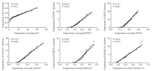
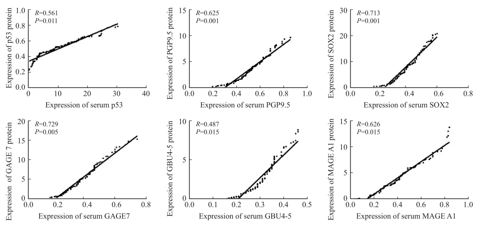
Fig. 1
Correlation analysis of tumor tissue protein expression and peripheral blood serum expression"
| [1] |
BADE B C, DELA CRUZ C S. Lung cancer 2020: epidemiology, etiology, and prevention[J]. Clin Chest Med, 2020, 41(1): 1-24.
doi: S0272-5231(19)30080-2 pmid: 32008623 |
| [2] |
ZHOU Y Y, HÖTI N, AO M H, et al. Expression of p16 and p53 in non-small cell lung cancer: clinicopathological correlation and potential prognostic impact[J]. Biomark Med, 2019, 13(9): 761-771.
doi: 10.2217/bmm-2018-0441 |
| [3] |
BRAIT M, MALDONADO L, NOORDHUIS M G, et al. Association of promoter methylation of VGF and PGP9.5 with ovarian cancer progression[J]. PLoS One, 2013, 8(9): e70878.
doi: 10.1371/journal.pone.0070878 |
| [4] |
WANG K X, JI W X, YU Y F, et al. FGFR1-ERK1/2-SOX2 axis promotes cell proliferation, epithelial-mesenchymal transition, and metastasis in FGFR1-amplified lung cancer[J]. Oncogene, 2018, 37(39): 5340-5354.
doi: 10.1038/s41388-018-0311-3 pmid: 29858603 |
| [5] |
WANG J, SHIVAKUMAR S, BARKER K, et al. Comparative study of autoantibody responses between lung adenocarcinoma and benign pulmonary nodules[J]. J Thorac Oncol, 2016, 11(3): 334-345.
doi: 10.1016/j.jtho.2015.11.011 pmid: 26896032 |
| [6] |
ORLOVETSKIE N, SERRUYA R, ABBOUD-JARROUS G, et al. Targeted inhibition of WRN helicase, replication stress and cancer[J]. Biochim Biophys Acta Rev Cancer, 2017, 1867(1): 42-48.
doi: 10.1016/j.bbcan.2016.11.004 |
| [7] |
YI E, CHANG J E, LEEM C, et al. Association of MAGE A1-6 expression with lung cancer progression[J]. J Cancer, 2017, 8(8): 1324-1329.
doi: 10.7150/jca.18086 pmid: 28638445 |
| [8] |
ZHANG X Z, LIU M, ZHANG X, et al. Autoantibodies to tumor-associated antigens in lung cancer diagnosis[J]. Adv Clin Chem, 2021, 103: 1-45.
doi: 10.1016/bs.acc.2020.08.005 pmid: 34229848 |
| [9] |
XIAO K J, MA X L, WANG Y C, et al. Diagnostic value of serum tumor-associated autoantibodies in esophageal cancer[J]. Biomark Med, 2021, 15(15): 1333-1343.
doi: 10.2217/bmm-2021-0351 pmid: 34541870 |
| [10] |
ZHANG R, MA L, LI W Y, et al. Diagnostic value of multiple tumor-associated autoantibodies in lung cancer[J]. Onco Targets Ther, 2019, 12: 457-469.
doi: 10.2147/OTT.S187734 |
| [11] |
DU Q, YU R F, WANG H, et al. Significance of tumor-associated autoantibodies in the early diagnosis of lung cancer[J]. Clin Respir J, 2018, 12(6): 2020-2028.
doi: 10.1111/crj.12769 pmid: 29356386 |
| [12] |
CHAPMAN C J, HEALEY G F, MURRAY A, et al. EarlyCDT®-Lung test: improved clinical utility through additional autoantibody assays[J]. Tumour Biol, 2012, 33(5): 1319-1326.
doi: 10.1007/s13277-012-0379-2 |
| [13] |
CHAPMAN C J, MURRAY A, MCELVEEN J E, et al. Autoantibodies in lung cancer: possibilities for early detection and subsequent cure[J]. Thorax, 2008, 63(3): 228-233.
doi: 10.1136/thx.2007.083592 pmid: 17932110 |
| [14] |
INFANTE M, CAVUTO S, LUTMAN F R, et al. A randomized study of lung cancer screening with spiral computed tomography: three-year results from the DANTE trial[J]. Am J Respir Crit Care Med, 2009, 180(5): 445-453.
doi: 10.1164/rccm.200901-0076OC |
| [15] |
PARRALES A, IWAKUMA T. Targeting oncogenic mutant p53 for cancer therapy[J]. Front Oncol, 2015, 5: 288.
doi: 10.3389/fonc.2015.00288 pmid: 26732534 |
| [16] |
QIN J Y, ZENG N, YANG T, et al. Diagnostic value of autoantibodies in lung cancer: a systematic review and meta-analysis[J]. Cell Physiol Biochem, 2018, 51(6): 2631-2646.
doi: 10.1159/000495935 pmid: 30562746 |
| [17] |
NING Y C, HUI N, QING B, et al. ZCCHC10 suppresses lung cancer progression and cisplatin resistance by attenuating MDM2-mediated p53 ubiquitination and degradation[J]. Cell Death Dis, 2019, 10(6): 414.
doi: 10.1038/s41419-019-1635-9 pmid: 31138778 |
| [18] |
SHAO L P, ZUO X L, YANG Y, et al. The inherited variations of a p53-responsive enhancer in 13q12.12 confer lung cancer risk by attenuating TNFRSF19 expression[J]. Genome Biol, 2019, 20(1): 103.
doi: 10.1186/s13059-019-1696-1 pmid: 31126313 |
| [19] |
REN S X, ZHANG S C, JIANG T, et al. Early detection of lung cancer by using an autoantibody panel in Chinese population[J]. Oncoimmunology, 2018, 7(2): e1384108.
doi: 10.1080/2162402X.2017.1384108 |
| [20] |
WEN W, LIU G, JIN K, et al. TGF-β1 induces PGP9.5 expression in CAFs to promote the growth of colorectal cancer cells[J]. Oncol Rep, 2017, 37(1): 115-122.
doi: 10.3892/or.2016.5238 pmid: 27840994 |
| [21] |
LIN S C, CHOU Y T, JIANG S S, et al. Epigenetic switch between SOX2 and SOX9 regulates cancer cell plasticity[J]. Cancer Res, 2016, 76(23): 7036-7048.
doi: 10.1158/0008-5472.CAN-15-3178 |
| [22] |
KAMEL L M, ATEF D M, MACKAWY A M H, et al. Circulating long non-coding RNA GAS5 and SOX2OT as potential biomarkers for diagnosis and prognosis of non-small cell lung cancer[J]. Biotechnol Appl Biochem, 2019, 66(4): 634-642.
doi: 10.1002/bab.1764 |
| [23] |
TANG Z M, LING Z G, WANG C M, et al. Serum tumor-associated autoantibodies as diagnostic biomarkers for lung cancer: a systematic review and meta-analysis[J]. PLoS One, 2017, 12(7): e0182117.
doi: 10.1371/journal.pone.0182117 |
| [24] | MU Y Y, XIE F Y, WANG F B, et al. Performance evaluation of an enzyme-linked immunosorbent assay for seven autoantibodies in lung cancer[J]. Clin Lab, 2019, 65(4). |
| [25] |
FANIPAKDEL A, SEILANIAN TOUSSI M, REZAZADEH F, et al. Overexpression of cancer-testis antigen melanoma-associated antigen A1 in lung cancer: a novel biomarker for prognosis, and a possible target for immunotherapy[J]. J Cell Physiol, 2019, 234(7): 12080-12086.
doi: 10.1002/jcp.27884 pmid: 30569450 |
| [1] | WANG Manli, CHEN Hui, DUAN Zhi, XU Qimei, LI Zhen. A study on communication mechanism of lung cancer cells in tumor microenvironment mediated by pleckstrin-2/miR-196a signal axis [J]. China Oncology, 2024, 34(7): 628-638. |
| [2] | QIAN Bin, CHEN Haiquan. Important progress in surgical treatment of lung cancer in 2023 [J]. China Oncology, 2024, 34(4): 335-339. |
| [3] | LIN Yicong, WANG Yue, XUE Qianqian, ZHENG Qiang, JIN Yan, HUANG Ziling, LI Yuan. Clinical pathological characteristics and immune microenvironment significance of EGFR T790M mutation in non-small cell lung cancer patients and its prognostic implications [J]. China Oncology, 2024, 34(4): 368-379. |
| [4] | JIANG Mengqi, HAN Yuchen, FU Xiaolong. Research progress on H-E stained whole slide image analysis by artificial intelligence in lung cancer [J]. China Oncology, 2024, 34(3): 306-315. |
| [5] | WU Han, YANG Zhangru, FENG Wen, ZENG Wanqin, GUO Jindong, LI Hongxuan, WANG Changlu, WANG Jiaming, LÜ Changxing, ZHANG Qin, YU Wen, CAI Xuwei, FU Xiaolong. The efficacy and prognosis analysis after stereotactic body radiotherapy for multiple primary early-stage lung cancer [J]. China Oncology, 2023, 33(9): 844-856. |
| [6] | ZHANG Lingling, WANG Xiangyi, WEI Xing, LIN Li, TANG Chuanhao, LIANG Jun. A study on prevention and treatment of chemotherapy induced nausea and vomiting in non-small cell lung cancer patients with low-frequency electrical stimulator for antiemesis [J]. China Oncology, 2023, 33(8): 776-781. |
| [7] | ZHANG Haoting, ZHENG Jing, FU Mengjiao, ZHOU Jianying. Research progress on thyroid dysfunction induced by immunotherapy for lung cancer [J]. China Oncology, 2023, 33(7): 701-706. |
| [8] | WU Jing, ZHOU Juan, SU Chunxia. Advances in fatty acid metabolism reprogramming of lung cancer [J]. China Oncology, 2023, 33(5): 517-526. |
| [9] | SU Chunxia, ZHOU Caicun. Important clinical research progress in lung cancer in 2022 [J]. China Oncology, 2023, 33(3): 218-227. |
| [10] | HE Liyuan, WANG Yudong. Research progress of ALK kinase domain drug resistance mutation and its future countermeasures [J]. China Oncology, 2022, 32(8): 736-746. |
| [11] | HONG Yaping, HUANG Yunjian, HUANG Zhangzhou, CHEN Shengjia, ZHONG Qiaofeng, ZENG Hongfu, ZHUANG Wu. Efficacy and prognostic predictors of first-generation EGFR TKI-targeted therapy in patients with EGFR-mutated advanced non-small cell lung cancer [J]. China Oncology, 2022, 32(7): 624-634. |
| [12] | WU Jianhui, CHU Xiangling, WANG Liqiang, LIN Xinqing, XIE Xiaohong, XIE Mengqing, ZHAO Jing, DENG Haiyi, YANG Yilin, QIU Guihuan, ZHOU Maolin, SUN Ni, LI Ru, CHEN Ying, DENG Jiaxi, ZENG Chen, PAN Bolin, QIN Yinyin, LIU Ming, SU Chunxia, ZHOU Chengzhi. Epidemiological analysis of real-world immune checkpoint inhibitor-related pneumonitis in Chinese patients with lung cancer [J]. China Oncology, 2022, 32(6): 469-477. |
| [13] | SU Chunxia, ZHOU Caicun. Current status and future directions of immunotherapy for advanced non-small cell lung cancer [J]. China Oncology, 2022, 32(6): 478-486. |
| [14] | YU Silai, NI Jianjiao, ZHU Zhengfei. Treatment of unresectable locally advanced non-small cell lung cancer in the era of immunotherapy: status and prospects [J]. China Oncology, 2022, 32(6): 487-498. |
| [15] | FU Yuanyuan, HOU Runping, FU Xiaolong. Research progress in predicting the risk of lymphatic or hematologic metastasis based on chest CT in early non-small cell lung cancer [J]. China Oncology, 2022, 32(4): 343-350. |
| Viewed | ||||||
|
Full text |
|
|||||
|
Abstract |
|
|||||
沪ICP备12009617
Powered by Beijing Magtech Co. Ltd