Welcome to China Oncology,
China Oncology ›› 2022, Vol. 32 ›› Issue (3): 234-242.doi: 10.19401/j.cnki.1007-3639.2022.03.006
• Article • Previous Articles Next Articles
WANG Zimao, CAO Yuan, WANG Qiying( )
)
Received:2021-10-27
Revised:2021-12-29
Online:2022-03-30
Published:2022-04-02
Contact:
WANG Qiying
E-mail:wangqiying@zzu.edu.cn
Share article
CLC Number:
WANG Zimao, CAO Yuan, WANG Qiying. Construction and validation of the survival prediction model for patients with cutaneous spindle cell melanoma[J]. China Oncology, 2022, 32(3): 234-242.
Tab. 1
Demographic and clinicopathological characteristics of 1 445 SCM patients [n (%)]"
| Characteristic | Total (n=1 445) | Training (n=1 011) | Validation (n=434) | P value |
|---|---|---|---|---|
| Age/year | 0.640 | |||
| ≤65 | 581 (40.2) | 411 (40.7) | 170 (39.2) | |
| ≥66 | 864 (59.8) | 600 (59.3) | 264 (60.8) | |
| Gender | 0.509 | |||
| Female | 483 (33.4) | 332 (32.8) | 151 (34.8) | |
| Male | 962 (66.6) | 679 (67.2) | 283 (65.2) | |
| Race | 0.786 | |||
| Non-white | 23 (1.6) | 15 (1.5) | 8 (1.8) | |
| White | 1422 (98.4) | 996 (98.5) | 426 (98.2) | |
| Site | 0.635 | |||
| Extremities | 557 (38.5) | 393 (38.9) | 164 (37.8) | |
| Scalp/face/neck | 600 (41.5) | 412 (40.8) | 188 (43.3) | |
| Trunk | 288 (20.0) | 206 (20.3) | 82 (18.9) | |
| Depth D/mm | 0.648 | |||
| ≤1.00 | 343 (23.7) | 238 (23.5) | 105 (24.2) | |
| 1.01-2.00 | 342 (23.7) | 243 (24.0) | 99 (22.8) | |
| 2.01-4.00 | 325 (22.5) | 234 (23.2) | 91 (21.0) | |
| ≥4.01 | 435 (30.1) | 296 (29.3) | 139 (32.0) | |
| Ulceration | 0.079 | |||
| Absent | 936 (64.8) | 670 (66.3) | 266 (61.3) | |
| Present | 509 (35.2) | 341 (33.7) | 168 (38.7) | |
| N stage | 0.779 | |||
| N0 | 1292 (89.4) | 904 (89.4) | 388 (89.4) | |
| N1 | 68 (4.7) | 45 (4.4) | 23 (5.3) | |
| N2 | 52 (3.6) | 39 (3.9) | 13 (3.0) | |
| N3 | 33 (2.3) | 23 (2.3) | 10 (2.3) | |
| M stage | 0.542 | |||
| M0 | 1412 (97.7) | 990 (97.9) | 422 (97.2) | |
| M1 | 33 (2.3) | 21 (2.1) | 12 (2.8) | |
| Surgery | 0.620 | |||
| No/unknown | 29 (2.0) | 22 (2.2) | 7 (1.6) | |
| Yes | 1416 (98.0) | 989 (97.8) | 427 (98.4) | |
| Radiotherapy | 0.329 | |||
| No/unknown | 1357 (93.9) | 954 (94.4) | 403 (92.9) | |
| Yes | 88 (6.1) | 57 (5.6) | 31 (7.1) | |
| Chemotherapy | 0.874 | |||
| No/unknown | 1412 (97.7) | 987 (97.6) | 425 (97.9) | |
| Yes | 33 (2.3) | 24 (2.4) | 9 (2.1) |
Tab. 2
Univariate COX regression analysis of SCM patients"
| Characteristic | CSS | OS | |||
|---|---|---|---|---|---|
| P value | HR (95% CI) | P value | HR (95% CI) | ||
| Age/year | |||||
| ≤65 | Ref | Ref | |||
| ≥66 | 0.000 | 2.30 (1.63-3.23) | 0.000 | 4.53 (3.54-5.81) | |
| Gender | |||||
| Female | Ref | Ref | |||
| Male | 0.015 | 1.54 (1.09-2.19) | 0.000 | 1.56 (1.25-1.93) | |
| Race | |||||
| Non-white | Ref | Ref | |||
| White | 0.318 | 0.60 (0.22-1.63) | 0.573 | 0.82 (0.41-1.65) | |
| Site | |||||
| Extremities | Ref | Ref | |||
| Scalp/face/neck | 0.000 | 1.98 (1.41-2.80) | 0.000 | 2.19 (1.76-2.71) | |
| Trunk | 0.961 | 0.99 (0.62-1.58) | 0.862 | 1.03 (0.77-1.37) | |
| Depth D/mm | |||||
| ≤1.00 | Ref | Ref | |||
| 1.01-2.00 | 0.190 | 1.51 (0.82-2.80) | 0.278 | 1.20 (0.86-1.66) | |
| 2.01-4.00 | 0.001 | 2.66 (1.50-4.70) | 0.000 | 2.07 (1.53-2.80) | |
| ≥4.01 | 0.000 | 5.47 (3.24-9.21) | 0.000 | 3.13 (2.37-4.15) | |
| N stage | |||||
| N0 | Ref | Ref | |||
| N1 | 0.000 | 2.86 (1.64-4.97) | 0.010 | 1.73 (1.14-2.61) | |
| N2 | 0.000 | 4.05 (2.36-6.93) | 0.001 | 2.12 (1.38-3.27) | |
| N3 | 0.000 | 12.04 (6.86-21.12) | 0.000 | 6.54 (4.09-10.44) | |
| M stage | |||||
| M0 | Ref | Ref | |||
| M1 | 0.000 | 6.04 (3.27-11.15) | 0.000 | 3.56 (2.16-5.88) | |
| Ulceration | |||||
| Absent | Ref | Ref | |||
| Present | 0.000 | 3.07 (2.26-4.17) | 0.000 | 2.59 (2.14-3.13) | |
| Surgery | |||||
| No/unknown | Ref | Ref | |||
| Yes | 0.032 | 0.41 (0.18-0.93) | 0.009 | 0.48 (0.28-0.83) | |
| Radiotherapy | |||||
| No/unknown | Ref | Ref | |||
| Yes | 0.039 | 1.78 (1.03-3.08) | 0.016 | 1.57 (1.09-2.26) | |
| Chemotherapy | |||||
| No/unknown | Ref | Ref | |||
| Yes | 0.000 | 4.82 (2.73-8.5) | 0.013 | 1.96 (1.15-3.34) | |
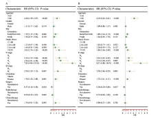
Fig. 1
Forest plot of SCM patients by multivariate COX regression analysis A: CSS; B: OS"
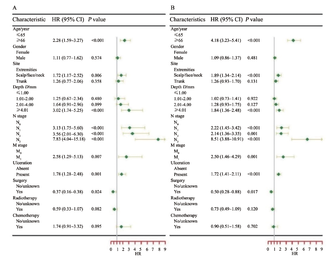
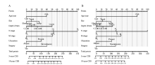
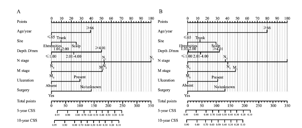
Fig. 2
Nomogram for predicting 5- and 10-year CSS (A) and OS (B) of patients with SCM"
Tab. 3
The corresponding score of each factor in the nomogram"
| Characteristic | Points (CSS) | Points (OS) |
|---|---|---|
| Age/year | ||
| ≤65 | 0 | 0 |
| ≥66 | 39 | 80 |
| Site | ||
| Extremities | 0 | 0 |
| Scalp/face/neck | 25 | 28 |
| Trunk | 10 | 12 |
| Depth D/mm | ||
| ≤1.00 | 0 | 0 |
| 1.01-2.00 | 10 | 1 |
| 2.01-4.00 | 22 | 13 |
| ≥4.01 | 50 | 32 |
| N stage | ||
| N0 | 0 | 0 |
| N1 | 53 | 40 |
| N2 | 53 | 38 |
| N3 | 100 | 100 |
| M stage | ||
| M0 | 0 | 0 |
| M1 | 51 | 48 |
| Ulceration | ||
| Absent | 0 | 0 |
| Present | 29 | 30 |
| Surgery | ||
| No/unknown | 40 | 38 |
| Yes | 0 | 0 |
Tab. 4
C-index of CSS and OS prediction models"
| Nomogram | Training cohorts C-index (95% CI) | Validation cohorts C-index (95% CI) |
|---|---|---|
| CSS | 0.778 (0.742-0.814) | 0.749 (0.696-0.802) |
| OS | 0.753 (0.731-0.775) | 0.712 (0.672-0.752) |
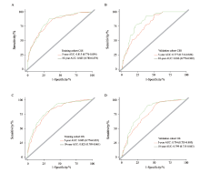
Fig. 3
ROC curves and AUC of 5- and10-year CSS (A and B) and OS (C and D) of SCM patients in the training cohort and validation cohort"
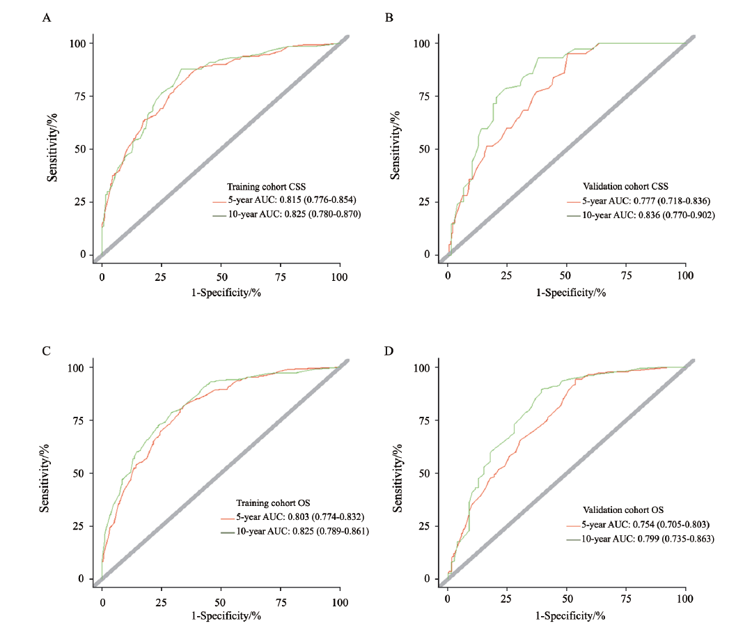
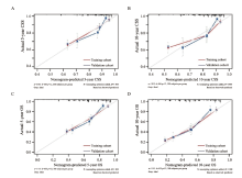
Fig. 4
Calibration curves for 5-year and 10-year CSS (A and B) and OS (C and D) predicted by nomogram in SCM patients"
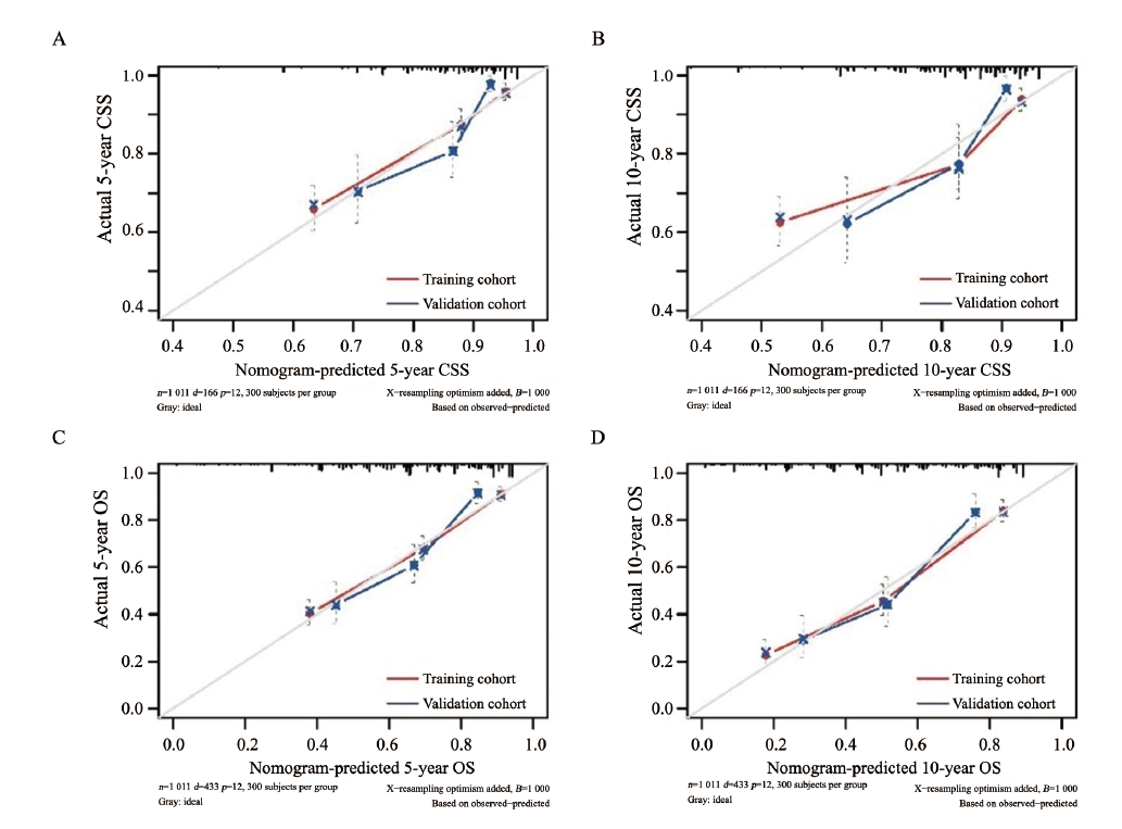
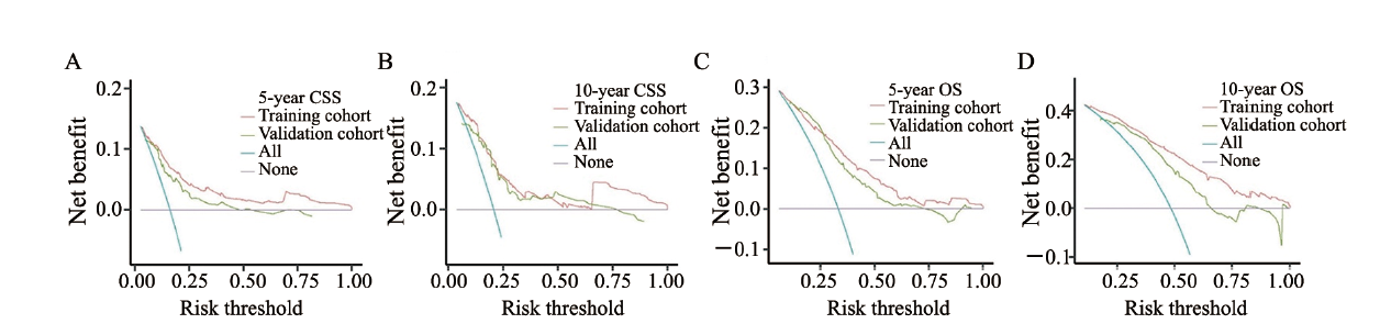
Fig. 5
DCA for training cohort and validation cohort to predict 5- and 10-year CSS (A and B) and OS (C and D) in SCM patients The abscissa represents threshold probability and the ordinate represents net benefit. The X-axis (purple line) shows that all samples are negative, and the net benefit is zero. The slash line (blue line) indicates that all samples are positive. The net benefit is expressed as a negative slope. The nomogram had the clinical net benefit in a wide range of threshold probabilities (0.10-0.99). A: Threshold probability of validation cohort (green line) was <0.44; B: Threshold probability of validation cohort (green line) was <0.75; C: Threshold probability of validation cohort (green line) was <0.67; D: Threshold probability of validation cohort (green line) was <0.65. DCA: Decision curve analysis."
| [1] | WINNEPENNINCKX V, DE VOS R, STAS M, et al. New phenotypical and ultrastructural findings in spindle cell (desmoplastic/neurotropic) melanoma[J]. Appl Immunohistochem Mol Morphol, 2003, 11(4): 319-325. |
| [2] |
WALIA R, JAIN D, MATHUR S R, et al. Spindle cell melanoma: a comparison of the cytomorphological features with the epithelioid variant[J]. Acta Cytol, 2013, 57(6): 557-561.
doi: 10.1159/000354405 |
| [3] |
KIM J, LAZAR A J, DAVIES M A, et al. BRAF, NRAS and KIT sequencing analysis of spindle cell melanoma[J]. J Cutan Pathol, 2012, 39(9): 821-825.
doi: 10.1111/j.1600-0560.2012.01950.x |
| [4] |
PIAO Y C, GUO M, GONG Y. Diagnostic challenges of metastatic spindle cell melanoma on fine-needle aspiration specimens[J]. Cancer, 2008, 114(2): 94-101.
doi: 10.1002/cncr.23345 |
| [5] |
BANERJEE S S, HARRIS M. Morphological and immunophenotypic variations in malignant melanoma[J]. Histopathology, 2000, 36(5): 387-402.
doi: 10.1046/j.1365-2559.2000.00894.x |
| [6] |
MORGAN M B, PUROHIT C, ANGLIN T R. Immunohistochemical distinction of cutaneous spindle cell carcinoma[J]. Am J Dermatopathol, 2008, 30(3): 228-232.
doi: 10.1097/DAD.0b013e31816de820 |
| [7] |
STOWMAN A M, MILLS S E, WICK M R. Spindle cell melanoma and interdigitating dendritic cell sarcoma: do they represent the same process?[J]. Am J Surg Pathol, 2016, 40(9): 1270-1279.
doi: 10.1097/PAS.0000000000000678 |
| [8] |
TACHA D, QI W M, RA S, et al. A newly developed mouse monoclonal SOX10 antibody is a highly sensitive and specific marker for malignant melanoma, including spindle cell and desmoplastic melanomas[J]. Arch Pathol Lab Med, 2015, 139(4): 530-536.
doi: 10.5858/arpa.2014-0077-OA |
| [9] | RAWANDALE N A, SURYAWANSHI K H. Primary spindle cell malignant melanoma of esophagus: an unusual finding[J]. J Clin Diagn Res, 2016, 10(2): OD03-OD04. |
| [10] |
DAINICHI T, KOBAYASHI C, FUJITA S, et al. Interdigital amelanotic spindle-cell melanoma mimicking an inflammatory process due to dermatophytosis[J]. J Dermatol, 2007, 34(10): 716-719.
doi: 10.1111/jde.2007.34.issue-10 |
| [11] | XU Z, SHI P, YIBULAYIN F, et al. Spindle cell melanoma: Incidence and survival, 1973-2017[J]. Oncol Lett, 2018, 16(4): 5091-5099. |
| [12] |
XU Z, YIBULAYIN F, SHI P, et al. Desmoplastic melanoma versus spindle cell melanoma: incidence and survival, 1973 to 2017[J]. Medicine (Baltimore), 2018, 97(29): e11563.
doi: 10.1097/MD.0000000000011563 |
| [13] |
GERSHENWALD J E, SCOLYER R A, HESS K R, et al. Melanoma staging: evidence-based changes in the American Joint Committee on Cancer eighth edition cancer staging manual[J]. CA Cancer J Clin, 2017, 67(6): 472-492.
doi: 10.3322/caac.21409 |
| [14] |
EL SHAROUNI M A, VAREY A H R, WITKAMP A J, et al. Predicting sentinel node positivity in patients with melanoma: external validation of a risk-prediction calculator (the Melanoma Institute Australia nomogram) using a large European population-based patient cohort[J]. Br J Dermatol, 2021, 185(2): 412-418.
doi: 10.1111/bjd.v185.2 |
| [15] |
MARCHETTI M A, LIOPYRIS K, NAVARRETE-DECHENT C. Net benefit and decision curve analysis of competing diagnostic strategies for cutaneous melanoma[J]. J Am Acad Dermatol, 2021, 85(2): e87-e88.
doi: 10.1016/j.jaad.2020.04.170 |
| [16] |
IASONOS A, SCHRAG D, RAJ G V, et al. How to build and interpret a nomogram for cancer prognosis[J]. J Clin Oncol, 2008, 26(8): 1364-1370.
doi: 10.1200/JCO.2007.12.9791 |
| [17] | WACHTEL J G, CAPLAN C W, MAKLEY T A Jr. Juvenile melanoma (mixed spindle cell and epithelioid cell nevus) of the conjunctiva[J]. Surv Ophthalmol, 1967, 12(1): 12-16. |
| [18] |
HOLLMIG S T, SACHDEV R, COCKERELL C J, et al. Spindle cell neoplasms encountered in dermatologic surgery: a review[J]. Dermatol Surg, 2012, 38(6): 825-850.
doi: 10.1111/j.1524-4725.2012.02296.x |
| [19] |
BALACHANDRAN V P, GONEN M, SMITH J J, et al. Nomograms in oncology: more than meets the eye[J]. Lancet Oncol, 2015, 16(4): e173-e180.
doi: 10.1016/S1470-2045(14)71116-7 |
| [20] |
GONG H Z, ZHENG H Y, LI J. Amelanotic melanoma[J]. Melanoma Res, 2019, 29(3): 221-230.
doi: 10.1097/CMR.0000000000000571 |
| [21] |
XIAO Y, PENG S S, HU Y H, et al. Development and validation of prognostic nomogram in patients with nonmetastatic malignant melanoma: a SEER population-based study[J]. Cancer Med, 2020, 9(22): 8562-8570.
doi: 10.1002/cam4.v9.22 |
| [22] |
VERVER D, VAN KLAVEREN D, FRANKE V, et al. Development and validation of a nomogram to predict recurrence and melanoma-specific mortality in patients with negative sentinel lymph nodes[J]. Br J Surg, 2019, 106(3): 217-225.
doi: 10.1002/bjs.10995 |
| [1] | HUANG Haozhe, CHEN Hong, ZHENG Dezhong, CHEN Chao, WANG Ying, XU Lichao, WANG Yaohui, HE Xinhong, YANG Yuanyuan, LI Wentao. A CT-based radiomics nomogram for predicting local tumor progression of colorectal cancer lung metastases treated with radiofrequency ablation [J]. China Oncology, 2024, 34(9): 857-872. |
| [2] | WU Wen, ZHANG Ruoxin, WENG Junyong, MA Yanlei, CAI Guoxiang, LI Xinxiang, YANG Yongzhi. Exploring the prognostic value of positive lymph node ratio in stage Ⅲ colorectal cancer patients and establishing a predictive model [J]. China Oncology, 2024, 34(9): 873-880. |
| [3] | XIAO Feng, XU Tonglin, ZHU Lin, XIAO Jingwen, WU Tianqi, GU Chunyan. Significance of infiltration of M1 tumor-associated macrophages in hepatocellular carcinoma [J]. China Oncology, 2024, 34(8): 726-733. |
| [4] | ZHANG Ruoxin, YE Zilan, WENG Junyong, LI Xinxiang. Correlation study between advanced age and inferior prognosis in stage Ⅱ colorectal cancer patients [J]. China Oncology, 2024, 34(5): 485-492. |
| [5] | SHEN Jie, WANG Jiangli, WANG Zezhou, MO Miao, ZHOU Changming, YUAN Jing, XU Dazhi, ZHENG Ying. Survival analysis of 6 737 surgically resected gastric cancer cases in China from a large single institution hospital-based cancer registry database [J]. China Oncology, 2024, 34(3): 268-277. |
| [6] | LI Jun, LU Tingwei, FANG Xuqian. Impact of MSI-H/dMMR on clinicopathological characteristics and prognosis of patients with BRAF V600E-mutated resectable colorectal cancer [J]. China Oncology, 2024, 34(11): 1061-1066. |
| [7] | JIN Yizi, LIN Mingxi, ZHANG Jian. Receptor discordance between primary breast cancer and liver metastases [J]. China Oncology, 2023, 33(9): 834-843. |
| [8] | WU Han, YANG Zhangru, FENG Wen, ZENG Wanqin, GUO Jindong, LI Hongxuan, WANG Changlu, WANG Jiaming, LÜ Changxing, ZHANG Qin, YU Wen, CAI Xuwei, FU Xiaolong. The efficacy and prognosis analysis after stereotactic body radiotherapy for multiple primary early-stage lung cancer [J]. China Oncology, 2023, 33(9): 844-856. |
| [9] | CHEN Jinjuan, WANG Xingran, LI Wenzhi, CHENG Yu, SUN Yihua, TAO Xiang, MA Fenghua, SUN Li, ZHAO Hongbo, LU Xin. Conservative surgery in stage I placental site trophoblastic tumor: a report of 10 cases and literature review [J]. China Oncology, 2023, 33(9): 857-865. |
| [10] | SUN Yang, WANG Lian, ZHAO Meng, ZHANG Xiaofeng, GENG Zhijun, WANG Yueyue, SONG Xue, ZUO Lugen, LI Jing, HU Jianguo. The prognostic value of high expression of FKBP1A in gastric cancer and the regulatory effect of targeted PI3K/AKT on glucose metabolism [J]. China Oncology, 2023, 33(8): 726-739. |
| [11] | JIANG Lin, LIU Qiying, JIA Liqing, ZHANG Jing, CHANG Heng, XUE Tian, REN Min, BAI Qianming, ZHU Xiaoli, ZHOU Xiaoyan. Retrospective study on MGMT methylation status and its clinical significance in gliomas [J]. China Oncology, 2023, 33(8): 740-750. |
| [12] | WANG Ruoxi, JI Peng, GONG Yue, CHEN Sheng. Response rate and clinical outcome of HER2-low breast cancer after neoadjuvant therapy: a single-center retrospective study [J]. China Oncology, 2023, 33(7): 686-692. |
| [13] | ZUO Xueliang, CHEN Zhiqiang, DONG Runyu, WANG Zhixiong, CAI Juan. The value of combined detection of LDHA and PD-L1 in predicting the efficacy and prognosis of advanced gastric cancer patients treated with PD-1 inhibitor [J]. China Oncology, 2023, 33(5): 460-468. |
| [14] | CHEN Yuguang, SUN Xiao, BI Zhao, QIU Pengfei, DUAN Baowei, FAN Qingda, WANG Yongsheng. Internal mammary sentinel lymph node biopsy for breast cancer: a long-term follow-up research for assessment of prognosis and guiding individualized internal mammary lymph node irradiation [J]. China Oncology, 2023, 33(2): 142-151. |
| [15] | CHEN Yingyao, CHU Xiangling, YU Xin, SU Chunxia. Advances in models predicting efficacy of immune checkpoint inhibitors [J]. China Oncology, 2023, 33(1): 61-70. |
| Viewed | ||||||
|
Full text |
|
|||||
|
Abstract |
|
|||||
沪ICP备12009617
Powered by Beijing Magtech Co. Ltd