Welcome to China Oncology,
China Oncology ›› 2023, Vol. 33 ›› Issue (3): 260-266.doi: 10.19401/j.cnki.1007-3639.2023.03.009
• Article • Previous Articles Next Articles
CHEN Ruchuan1,2( ), LIU Wei2, ZHOU Bingni2, LIU Xiaohang2, ZHOU Liangping1,2()
Received:2022-07-15
Revised:2023-02-27
Online:2023-03-30
Published:2023-04-17
Contact:
ZHOU Liangping
Share article
CLC Number:
CHEN Ruchuan, LIU Wei, ZHOU Bingni, LIU Xiaohang, ZHOU Liangping. The value of VI-RADS combined with tumor contact length in the detection of muscle-invasive bladder cancer[J]. China Oncology, 2023, 33(3): 260-266.
Tab. 1
MRI sequences and related parameters"
| Parameter | T1WI (axial) | T2WI (axial) | T2WI (sagittal) | DWI (axial) | DCE (axial) |
|---|---|---|---|---|---|
| Repetition time/ms | 231.00 | 1 500.00 | 6 760.00 | 6 210.00 | 3.56 |
| Echo time/ms | 2.46 | 101.00 | 101.00 | 62.00 | 1.39 |
| Slice thickness/mm | 5.5 | 1.5 | 4.0 | 4.0 | 3.0 |
| Slice interval/mm | 4 | 1 | 1 | 1 | 1 |
| Field of vision (mm×mm) | 300×300 | 300×300 | 230×230 | 320×320 | 330×200 |
| B value/(s·mm-2) | / | / | / | 0, 1 000 | / |
Tab. 2
VI-RADS score and pathology examination results of target lesions"
| VI-RADS | 1 | 2 | 3A | 3B | 4 | 5 | Total |
|---|---|---|---|---|---|---|---|
| NMIBC | 10 | 45 | 13 | 3 | 4 | 0 | 75 |
| MIBC | 0 | 9 | 3 | 22 | 22 | 28 | 84 |
Fig. 1
MRI image and outline diagram of classic case 1 A 55-year-old man with a bladder tumor underwent multiparametric MRI before primary TURBT. Pathology findings showed that urothelial cancer cells infiltrate only the mucosal layer. A: T2WI shows a broad-based tumor without high signal intensity thickened inner layer but with no clear disruption of low signal intensity muscularis on the right-lateral wall. VI-RADS score for T2W imaging was 3. B: DWI (b = 1 000) shows a broad-based tumor with restricted diffusion but with no clear disruption of low signal intensity muscularis propria. VI-RADS score for DWI was 3. C: DCE image shows early enhancement of the muscularis propria with no clear disruption of low signal intensity muscularis propria. VI-RADS score for DCE was 3. D: After measurement, the TCL of tumor reached a maximum of 2.5 cm on the axial view of T2WI and was classified to to VI-RADS 3A."
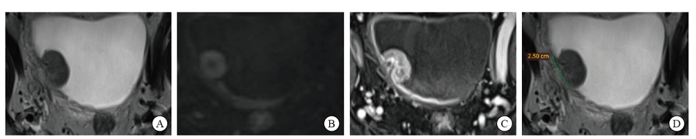
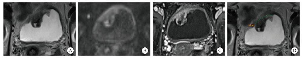
Fig. 2
MRI image and outline diagram of classic case 2 A 69-year-old woman with a bladder tumor underwent multiparametric MRI before primary TURBT. Pathology findings showed that there was urothelial carcinoma cell infiltration in the muscle layer. A: T2WI shows a broad-based tumor without high signal intensity thickened inner layer but with no clear disruption of low signal intensity muscularis on the right-lateral wall. VI-RADS score for T2W imaging was 3. B: DWI (b = 1000) shows a broad-based tumor with restricted diffusion but with no clear disruption of low signal intensity muscularis propria. VI-RADS score for DWI was 3. C: DCE image shows early enhancement of the muscularis propria with no clear disruption of low signal intensity muscularis propria. VI-RADS score for DCE was 3. D: After measurement, the TCL of tumor reached a maximum of 4.23 cm on the axial view of T2WI and was classified to VI-RADS 3B."
Tab. 3
Accuracy analysis of various diagnostic models in assessing MIBC"
| Assessment | Sensitivity/% | Specificity/% | PPV/% | NPV/% | Accuracy/% | Youden index |
|---|---|---|---|---|---|---|
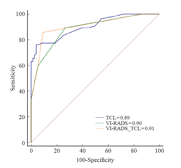
| VI-RADS (3) | 89.34 | 73.31 | 78.96 | 85.93 | 81.89 | 0.63 |
| VI-RADS (4) | 59.52 | 94.67 | 92.59 | 67.62 | 76.10 | 0.54 |
| TCL | 77.38 | 81.33 | 82.27 | 76.25 | 79.24 | 0.59 |
| VI-RADS_TCL | 85.73 | 90.72 | 91.11 | 85.07 | 88.19 | 0.76 |
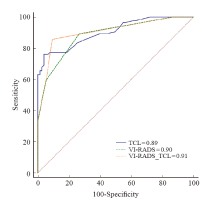
Fig. 3
ROC curve of TCL, VI-RADS and VI-RADS_TLC for predicting MIBC"
| [1] |
SIEGEL R L, MILLER K D, JEMAL A. Cancer statistics, 2015[J]. CA Cancer J Clin, 2015, 65(1): 5-29.
doi: 10.3322/caac.21254 |
| [2] |
PATEL V G, OH W K, GALSKY M D. Treatment of muscle-invasive and advanced bladder cancer in 2020[J]. CA Cancer J Clin, 2020, 70(5): 404-423.
doi: 10.3322/caac.v70.5 |
| [3] |
WITJES J A, BRUINS H M, CATHOMAS R, et al. European association of urology guidelines on muscle-invasive and metastatic bladder cancer: summary of the 2020 guidelines[J]. Eur Urol, 2021, 79(1): 82-104.
doi: 10.1016/j.eururo.2020.03.055 pmid: 32360052 |
| [4] |
THOENY H C, BELLIN M F, COMPERAT E M, et al. Vesical imaging-reporting and data system (VI-RADS): added value for management of bladder cancer patients?[J]. Eur Urol, 2018, 74(3): 307-308.
doi: S0302-2838(18)30442-1 pmid: 29960749 |
| [5] |
MENG X Y, HU H L, WANG Y C, et al. Accuracy and challenges in the vesical imaging-reporting and data system for staging bladder cancer[J]. J Magn Reson Imaging, 2022, 56(2): 391-398.
doi: 10.1002/jmri.v56.2 |
| [6] |
WANG H J, LUO C, ZHANG F, et al. Multiparametric MRI for bladder cancer: validation of VI-RADS for the detection of detrusor muscle invasion[J]. Radiology, 2019, 291(3): 668-674.
doi: 10.1148/radiol.2019182506 pmid: 31012814 |
| [7] |
GREEN R W, EPSTEIN E. Dynamic contrast-enhanced ultrasound improves diagnostic performance in endometrial cancer staging[J]. Ultrasound Obstet Gynecol, 2020, 56(1): 96-105.
doi: 10.1002/uog.21885 pmid: 31647145 |
| [8] |
LIU Y, LIU H, QIAN C L, et al. Utility of quantitative contrast-enhanced ultrasound for the prediction of extracapsular extension in papillary thyroid carcinoma[J]. Sci Rep, 2017, 7(1): 1472.
doi: 10.1038/s41598-017-01650-2 pmid: 28469180 |
| [9] |
FENG S H, YANG S T. The new 8th TNM staging system of lung cancer and its potential imaging interpretation pitfalls and limitations with CT image demonstrations[J]. Diagn Interv Radiol, 2019, 25(4): 270-279.
doi: 10.5152/dir.2019.18458 pmid: 31295144 |
| [10] |
BABJUK M, BURGER M, COMPÉRAT E M, et al. European Association of Urology guidelines on non-muscle-invasive bladder cancer (TaT1 and carcinoma in situ) -2019 update[J]. Eur Urol, 2019, 76(5): 639-657.
doi: 10.1016/j.eururo.2019.08.016 |
| [11] |
AHN H, HWANG S I, LEE H J, et al. Quantitation of bladder cancer for the prediction of muscle layer invasion as a complement to the vesical imaging-reporting and data system[J]. Eur Radiol, 2021, 31(3): 1656-1666.
doi: 10.1007/s00330-020-07224-7 |
| [12] |
PANEBIANCO V, NARUMI Y, ALTUN E, et al. Multiparametric magnetic resonance imaging for bladder cancer: development of VI-RADS (vesical imaging-reporting and data system)[J]. Eur Urol, 2018, 74(3): 294-306.
doi: S0302-2838(18)30335-X pmid: 29755006 |
| [13] |
PANER G P, STADLER W M, HANSEL D E, et al. Updates in the eighth edition of the tumor-node-metastasis staging classification for urologic cancers[J]. Eur Urol, 2018, 73(4): 560-569.
doi: S0302-2838(17)31064-3 pmid: 29325693 |
| [14] |
BARCHETTI G, SIMONE G, CERAVOLO I, et al. Multiparametric MRI of the bladder: inter-observer agreement and accuracy with the Vesical Imaging-Reporting and Data System (VI-RADS) at a single reference center[J]. Eur Radiol, 2019, 29(10): 5498-5506.
doi: 10.1007/s00330-019-06117-8 pmid: 30887202 |
| [15] | 保文斌, 栾婷, 郝金钢, 等. VI-RADS在膀胱癌中运用的研究进展及其局限性[J]. 医学影像学杂志, 2022, 32(5): 849-852. |
| BAO W B, LUAN T, HAO J G, et al. Research progress and limitations of VI-RADS in bladder cancer[J]. J Med Imaging, 2022, 32(5): 849-852. | |
| [16] | 中国肿瘤医院泌尿肿瘤协作组. 中国膀胱癌保膀胱治疗多学科诊治协作共识[J]. 中华肿瘤杂志, 2022, 44(3): 209-218. |
| China Cancer Hospital urinary Cancer Collaboration Group. Expert consensus of multi-disciplinary collaboration on bladder-preserving treatment for bladder cancer in China[J]. Chin J Oncol, 2022, 44(3): 209-218. | |
| [17] | MAKBOUL M, FARGHALY S, ABDELKAWI I F. Multiparametric MRI in differentiation between muscle invasive and non-muscle invasive urinary bladder cancer with vesical imaging reporting and data system (VI-RADS) application[J]. Br J Radiol, 2019, 92(1104): 20190401. |
| [18] |
MARCHIONI M, PRIMICERI G, DELLI PIZZI A, et al. Could bladder multiparametric MRI be introduced in routine clinical practice? Role of the new VI-RADS score: results from a prospective study[J]. Clin Genitourin Cancer, 2020, 18(5): 409-415.e1.
doi: 10.1016/j.clgc.2020.03.002 |
| [19] |
WANG X Y, TU N, SUN F, et al. Detecting muscle invasion of bladder cancer using a proposed magnetic resonance imaging strategy[J]. J Magn Reson Imaging, 2021, 54(4): 1212-1221.
doi: 10.1002/jmri.27676 pmid: 33998725 |
| Viewed | ||||||
|
Full text |
|
|||||
|
Abstract |
|
|||||
沪ICP备12009617
Powered by Beijing Magtech Co. Ltd