Welcome to China Oncology,
China Oncology ›› 2022, Vol. 32 ›› Issue (2): 106-117.doi: 10.19401/j.cnki.1007-3639.2022.02.002
Previous Articles Next Articles
ZHAO Jiaxian1,2, JIANG Daohuai3,4,5, CONG Binbin1,2, GAO Fei3,4( ), WANG Yongsheng1,2()
Received:2021-12-24
Revised:2022-01-10
Online:2022-02-28
Published:2022-03-08
Contact:
GAO Fei, WANG Yongsheng
E-mail:gaofei@shanghaitech.edu.cn;wangysh2008@aliyun.com
Share article
CLC Number:
ZHAO Jiaxian, JIANG Daohuai, CONG Binbin, GAO Fei, WANG Yongsheng. Basic research on photoacoustic sensing and imaging system for detecting sentinel lymph node in breast cancer[J]. China Oncology, 2022, 32(2): 106-117.
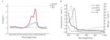
Fig. 1
Comparison of optical absorption spectra of different photoacoustic agents A: The absorption peaks of ICG and ICG-RIT were 780-800 nm; B: The light absorption peaks of methylene blue, methemoglobin, oxyhemoglobin and deoxyhemoglobin were located at 660-680 nm, 620-640 nm and below 600 nm, respectively."
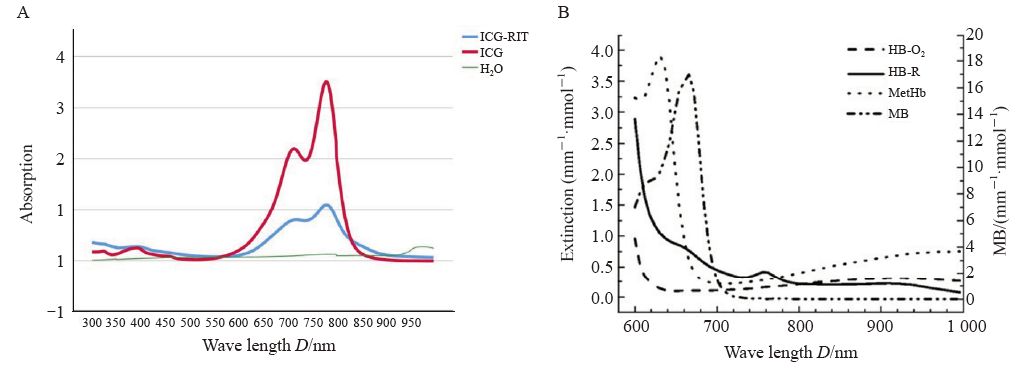
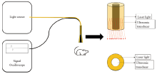
Fig.2
The diagram of the structure of hand-held photoacoustic signal sensing system"

Fig. 3
The structure of hand-held photoacoustic imaging system"

Tab. 1
Amplitudes of ICG-RIT phantoms with different concentrations at different depths"
| Depth D/mm | PA intensity of different agents U/mV | MB | Chicken breast tissue | ||||
|---|---|---|---|---|---|---|---|
| 100%ICG-RIT | 50%ICG-RIT | 25%ICG-RIT | 12.5%ICG-RIT | 6.25%ICG-RIT | |||
| 0.00 | 26.70 | 20.99 | 12.60 | 9.79 | 9.51 | 19.45 | 0 |
| 15.13 | 15.80 | 12.04 | 9.66 | 9.23 | 6.99 | 11.06 | |
| 24.84 | 13.58 | 8.39 | 7.84 | 7.41 | 6.17 | 9.93 | |
| 39.72 | 10.22 | 7.56 | 6.85 | 4.61 | 3.78 | 5.73 | |
| 52.42 | 5.87 | 4.62 | 3.42 | 3.36 | 0.00 | 4.34 | |
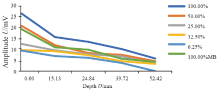
Fig. 4
Amplitudes of ICG-RIT phantoms with different concentrations at different depths"
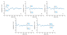
Fig. 5
Photoacoustic signals of 25% concentration ICG-RIT phantoms at different depths A: PA signal amplitude was 11.20 mV; Depth was 6.15 mm; B: PA signal amplitude was 8.99 mV; Depth was 17.37 mm; C: PA signal amplitude was 7.00 mV; Depth was 28.56 mm; D: PA signal amplitude was 6.25 mV; Depth was 43.50 mm; E: PA signal amplitude was 2.75 mV; Depth was 62.22 mm."
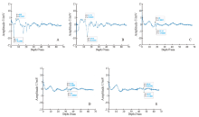
Fig. 6
Photoacoustic signals of 6.25% concentration ICG-RIT phantoms at different depths A: PA signal amplitude was 13.24 mV; Depth was 6.81 mm; B: PA signal amplitude was 12.25 mV; Depth was 14.73 mm; C: PA signal amplitude was 5.00 mV; Depth was 19.14 mm; D: PA signal amplitude was 2.74 mV; Depth was 33.00 mm; E: Photoacoustic signal at 46.95 mm depth was not used because of the low differentiation."
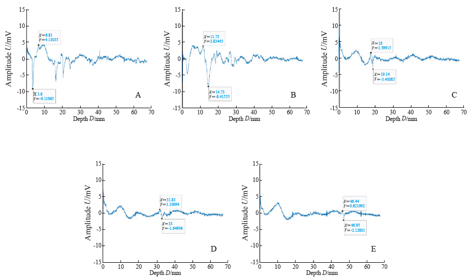
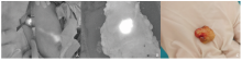
Fig.7
Lymph nodes in NIR fluorescence imaging system A: Lymph drainage in the NIR fluorescence imaging system after subareolar injection of ICG-RIT; B: Image of excised SLN in the NIR fluorescence imaging system; C:Visual inspection of the excised SLN."

Fig. 8
Photoacoustic signals SLN at different depths A: The photoacoustic signal of axillary fat; B: PA signal amplitude was 99 mV; Depth was 9.18 mm; C: PA signal amplitude was 72 mV; Depth: 17.37 mm; D: PA signal amplitude was 59 mV; Depth was 25.60 mm; E: PA signal amplitude was 31 mV; Depth was 32.72 mm."
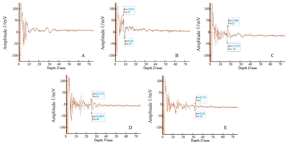
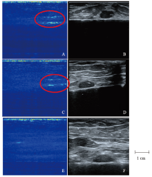
Fig. 9
Photoacoustic images and ultrasound images of SLN at different depths A, B: Both PAI and US imaged SLN under about 1 cm thick axillary fat; C, D: Both PAI and US imaged SLN under about 2.5 cm thick axillary fat; E: No significant SLN signal was found in PAI under about 4 cm thick axillary fat; F: US imaged SLN under about 4 cm thick axillary fat."


Fig. 10
Amplitudes and photoacoustic images after injection of ICG-RIT A, B: No significant signal of the popliteal LNs in the PASS and PAI was observed with no ICG-RIT injection; C, D: Characteristic photoacoustic signal in the PASS and PAI of the popliteal LNs was detected at 10.05 mm depth after ICG-RIT injection; E, F: No significant signal of the iliac LNs in the PASS and PAI was observed after ICG-RIT injection; G, H: No obvious staining of the popliteal LNs and the iliac LNs dissected along photoacoustic signal."

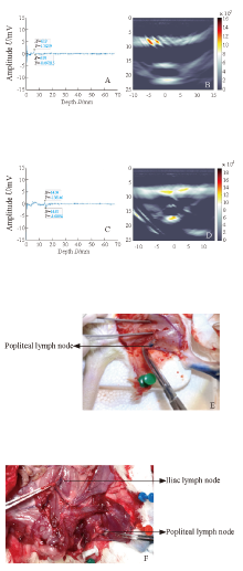
Fig. 11
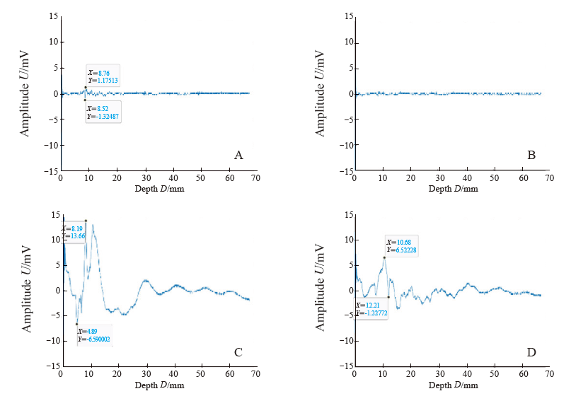
Amplitudes and photoacoustic images after injection of MB A, B: Characteristic photoacoustic signal in the PASS and PAI of the popliteal LNs was detected at 6.93 mm depth after MB injection; C, D: Characteristic photoacoustic signal in the PASS and PAI of the iliac LNs was detected at 14.67 mm depth after MB injection; E, F: Obvious staining of the popliteal LNs and the iliac LNs dissected along photoacoustic signal."

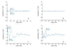
Fig. 12
Amplitudes of ex vivo lymph nodes A: ICG-RIT injection ex vivo popliteal LNs PA signal amplitude was 2.50 mV; B: ICG-RIT injection ex vivo popliteal LNs PA signal amplitude was 0 mV; C: MB injection ex vivo popliteal LNs PA signal amplitude was 20.25 mV; D: MB injection ex vivo popliteal LNs PA signal amplitude was 7.74 mV."
Tab. 2
Amplitude differences between ICG-RIT and MB at different level lymph nodes"
| Item | In vivo | Ex vivo | ||||
|---|---|---|---|---|---|---|
| Contralateral popliteal lymph node | Popliteal lymph node | Iliac lymph node | Popliteal lymph node | Iliac lymph node | ||
| ICG-RIT | 0.00 | 2.00 | 0.00 | 2.50 | 0.00 | |
| MB | 0.00 | 2.25 | 3.00 | 20.25 | 7.74 | |
| [1] | PARK Y H,, SENKUS-KONEFKA E,, IM S A, et al. Pan-Asian adapted ESMO Clinical Practice Guidelines for the management of patients with early breast cancer: a KSMO-ESMO initiative endorsed by CSCO, ISMPO, JSMO, MOS, SSO and TOS[J]. Ann Oncol, 2020,31(4):451-469. |
| [2] | GRADISHAR W J,, ANDERSON B O,, ABRAHAM J, et al. Breast cancer, version 3. 2020, NCCN clinical practice guidelines in oncology[J]. J Natl Compr Canc Netw, 2020,18(4):452-478. |
| [3] | GIULIANO A E,, BALLMAN K V,, MCCALL L, et al. Effect of axillary dissection vs no axillary dissection on 10-year overall survival among women with invasive breast cancer and sentinel node metastasis: the ACOSOG Z0011 (alliance) randomized clinical trial[J]. JAMA, 2017,318(10):918-926. |
| [4] | LI J Y,, CHEN X,, QI M, et al. Sentinel lymph node biopsy mapped with methylene blue dye alone in patients with breast cancer: a systematic review and meta-analysis[J]. PLoS One, 2018,13(9):e0204364. |
| [5] | RUBIO I T,, DIAZ-BOTERO S,, ESGUEVA A, et al. The superparamagnetic iron oxide is equivalent to the Tc99 radiotracer method for identifying the sentinel lymph node in breast cancer[J]. Eur J Surg Oncol, 2015,41(1):46-51. |
| [6] | CONG B B,, SUN X,, SONG X R, et al. Preparation study of indocyanine green-rituximab: a new receptor-targeted tracer for sentinel lymph node in breast cancer[J]. Oncotarget, 2016,7(30):47526-47535. |
| [7] | 丛斌斌,, 刘治国,, 孙晓, 等. 新型荧光靶向前哨淋巴结示踪剂的验证研究[J]. 中国癌症杂志, 2020,30(3):179-185. |
| CONG B B,, LIU Z G,, SUN X, et al. The validation study of a new fluorescence-target tracer for sentinel lymph node biopsy[J]. China Oncol, 2020,30(3):179-185. | |
| [8] | 赵家贤,, 王春建,, 丛斌斌, 等. 乳腺癌前哨淋巴结活检光声示踪剂的进展与展望[J]. 中国癌症杂志, 2021,31(10):873-878. |
| ZHAO J X,, WANG C J,, CONG B B, et al. Research progress of photoacoustic imaging in sentinel lymph node biopsy in breast cancer[J]. China Oncol, 2021,31(10):873-878. | |
| [9] | WEBER J,, BEARD P C,, BOHNDIEK S E. Contrast agents for molecular photoacoustic imaging[J]. Nat Methods, 2016,13(8):639-650. |
| [10] | KIM C,, SONG K H,, GAO F, et al. Sentinel lymph nodes and lymphatic vessels: noninvasive dual-modality in vivo mapping by using indocyanine green in rats: volumetric spectroscopic photoacoustic imaging and planar fluorescence imaging[J]. Radiology, 2010,255(2):442-450. |
| [11] | LEE J,, EL-ABADDI N,, DUKE A, et al. Noninvasive in vivo monitoring of methemoglobin formation and reduction with broadband diffuse optical spectroscopy[J]. J Appl Physiol (1985), 2006,100(2):615-622. |
| [12] | TROMBERG B J,, COQUOZ O,, FISHKIN J B, et al. Non-invasive measurements of breast tissue optical properties using frequency-domain photon migration[J]. Philos Trans R Soc Lond B Biol Sci, 1997,352(1354):661-668. |
| [13] | MARQUEZ G,, WANG L V,, LIN S P, et al. Anisotropy in the absorption and scattering spectra of chicken breast tissue[J]. Appl Opt, 1998,37(4):798-804. |
| [14] | 韩建红,, 甘仲霖,, 李达兵, 等. 三种鼠淋巴引流特点的比较研究[J]. 泸州医学院学报, 2015,38(4):388-392. |
| HAN J H,, GAN Z L,, LI D B, et al. Comparative study of lymphatic drainage patterns on three species of murine[J]. J Luzhou Med Coll, 2015,38(4):388-392. | |
| [15] | 中国抗癌协会乳腺癌专业委员会. 中国抗癌协会乳腺癌诊治指南与规范(2021年版)[J]. 中国癌症杂志, 2021,31(10):954-1040. |
| The Society of Breast Cancer China Anti-Cancer Association. Guidelines for breast cancer diagnosis and treatment by China Anti-Cancer Association (2021 edition)[J]. China Oncol, 2021,31(10):954-1040. | |
| [16] | TIAN C L,, SUN X,, CONG B B, et al. Murine model study of a new receptor-targeted tracer for sentinel lymph node in breast cancer[J]. J Breast Cancer, 2019,22(2):274-284. |
| [1] | XU Rui, WANG Zehao, WU Jiong. Advances in the role of tumor-associated neutrophils in the development of breast cancer [J]. China Oncology, 2024, 34(9): 881-889. |
| [2] | CAO Xiaoshan, YANG Beibei, CONG Binbin, LIU Hong. The progress of treatment for brain metastases of triple-negative breast cancer [J]. China Oncology, 2024, 34(8): 777-784. |
| [3] | ZHANG Jian. Clinical consideration of two key questions in assessing menopausal status of female breast cancer patients [J]. China Oncology, 2024, 34(7): 619-627. |
| [4] | JIANG Dan, SONG Guoqing, WANG Xiaodan. Study on the mechanism of mitochondrial dysfunction and CPT1A/ERK signal transduction pathway regulating malignant behavior in breast cancer [J]. China Oncology, 2024, 34(7): 650-658. |
| [5] | DONG Jianqiao, LI Kunyan, LI Jing, WANG Bin, WANG Yanhong, JIA Hongyan. A study on mechanism of SIRT3 inducing endocrine drug resistance in breast cancer via deacetylating YME1L1 [J]. China Oncology, 2024, 34(6): 537-547. |
| [6] | HAO Xian, HUANG Jianjun, YANG Wenxiu, LIU Jinting, ZHANG Junhong, LUO Yubei, LI Qing, WANG Dahong, GAO Yuwei, TAN Fuyun, BO Li, ZHENG Yu, WANG Rong, FENG Jianglong, LI Jing, ZHAO Chunhua, DOU Xiaowei. Establishment of primary breast cancer cell line as new model for drug screening and basic research [J]. China Oncology, 2024, 34(6): 561-570. |
| [7] | Committee of Breast Cancer Society, China Anti-Cancer Association. Expert consensus on clinical applications of ovarian function suppression for Chinese women with early breast cancer (2024 edition) [J]. China Oncology, 2024, 34(3): 316-333. |
| [8] | ZHANG Qi, XIU Bingqiu, WU Jiong. Progress of important clinical research of breast cancer in China in 2023 [J]. China Oncology, 2024, 34(2): 135-142. |
| [9] | ZHANG Siyuan, JIANG Zefei. Important research progress in clinical practice for advanced breast cancer in 2023 [J]. China Oncology, 2024, 34(2): 143-150. |
| [10] | WANG Zhaobu, LI Xing, YU Xinmiao, JIN Feng. Important research progress in clinical practice for early breast cancer in 2023 [J]. China Oncology, 2024, 34(2): 151-160. |
| [11] | LUO Yang, SUN Tao, SHAO Zhimin, CUI Jiuwei, PAN Yueyin, ZHANG Qingyuan, CHENG Ying, LI huiping, YANG Yan, YE Changsheng, YU Guohua, WANG Jingfen, LIU Yunjiang, LIU Xinlan, ZHOU Yuhong, BAI Yuju, GU Yuanting, WANG Xiaojia, XU Binghe, SONG Lihua. Efficacy, metabolic characteristics, safety and immunogenicity of AK-HER2 compared with reference trastuzumab in patients with metastatic HER2-positive breast cancer: a multicenter, randomized, double-blind phase Ⅲ equivalence trial [J]. China Oncology, 2024, 34(2): 161-175. |
| [12] | CHEN Yuanxiang, YU Tao, YANG Shiyu, ZENG Tao, WEI Lan, ZHANG Yan. KDM4A promotes the migration and invasion of breast cancer cell line MDA-MB-231 by downregulating BMP9 [J]. China Oncology, 2024, 34(2): 176-184. |
| [13] | HU Xiaoyu, CAI Yuwen, YE Fugui, SHAO Zhimin, HU Weigang, YU Keda. Impact of BRCA1/2 germline mutation on the incidence of second primary cancer following postoperative radiotherapy in patients with triple-negative breast cancer [J]. China Oncology, 2024, 34(2): 185-190. |
| [14] | ZHANG Siwei, MA Ding, JIANG Yizhou, SHAO Zhimin. “Subtype-precise” therapy leads diagnostic and therapeutic innovations: a new pattern for precision treatment of breast cancer [J]. China Oncology, 2024, 34(11): 1045-1052. |
| [15] | OUYANG Fei, WANG Yang, CHEN Yu, PEI Guoqing, WANG Ling, ZHANG Yang, SHI Lei. Construction of the prediction model of breast cancer bone metastasis based on machine learning [J]. China Oncology, 2024, 34(10): 903-914. |
| Viewed | ||||||
|
Full text |
|
|||||
|
Abstract |
|
|||||
沪ICP备12009617
Powered by Beijing Magtech Co. Ltd