Welcome to China Oncology,
China Oncology ›› 2022, Vol. 32 ›› Issue (3): 207-217.doi: 10.19401/j.cnki.1007-3639.2022.03.003
• Article • Previous Articles Next Articles
ZHU Haipeng1, HU Jun1, JIANG Min2, CAI Ruonan1, WANG Junqiao1, LI Li1( )
)
Received:2021-09-13
Revised:2021-12-06
Online:2022-03-30
Published:2022-04-02
Contact:
LI Li
E-mail:347993446@qq.com
Share article
CLC Number:
ZHU Haipeng, HU Jun, JIANG Min, CAI Ruonan, WANG Junqiao, LI Li. A study on mechanism of GOLM1 regulating PI3K/AKT/mTOR signaling pathway to promote proliferation, invasion and migration of lung adenocarcinoma cells[J]. China Oncology, 2022, 32(3): 207-217.
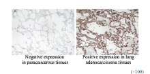
Fig. 1
Immunohistochemical staining to detect GOLM1 expression"
Tab. 1
Relationship between GOLM1 expression and clinicopathological characteristics in lung adenocarcinoma tissues [n (%)]"
| Characteristic | Case n | GOLM1 | χ2 | P value | |
|---|---|---|---|---|---|
| Negative | Positive | ||||
| Gender | 0.436 | 0.509 | |||
| Male | 47 | 9 (19.15) | 38 (80.85) | ||
| Female | 43 | 6 (13.95) | 37 (86.05) | ||
| Age/year | 0.009 | 0.925 | |||
| <60 | 41 | 7 (17.07) | 34 (82.93) | ||
| ≥60 | 49 | 8 (16.33) | 41 (83.67) | ||
| Smoking | 0.010 | 0.921 | |||
| Yes | 31 | 5 (16.13) | 26 (83.87) | ||
| No | 59 | 10 (16.95) | 49 (83.05) | ||
| Degree of differentiation | 9.681 | 0.002 | |||
| Medium/low differentiation | 56 | 4 (7.14) | 52 (92.86) | ||
| High differentiation | 34 | 11 (32.35) | 23 (67.65) | ||
| Lymph node metastasis | 21.056 | <0.001 | |||
| Yes | 34 | 8 (23.53) | 26 (76.47) | ||
| No | 56 | 41 (73.21) | 15 (26.79) | ||
| Clinical stage | 6.828 | 0.009 | |||
| Ⅰ/Ⅱ | 59 | 28 (47.46) | 31 (52.54) | ||
| Ⅲ | 31 | 6 (19.35) | 25 (80.65) | ||
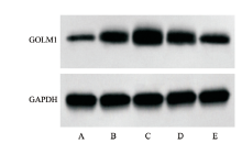
Fig. 2
Detection of GOLM1 expression in each group of cells by Western blot A: BEAS-2B cell; B: H460 cell; C: A549 cell; D: PG49 cell; E: H1299 cell."
Tab. 2
Comparison of the relative expression levels of GOLM1 in each group of cells ($\bar{x}±s$, n=6)"
| Group | GOLM1/GAPDH |
|---|---|
| BEAS-2B cell | 0.17±0.02 |
| H460 cell | 0.95±0.06a |
| A549 cell | 1.53±0.13a |
| PG49 cell | 1.13±0.10a |
| H1299 cell | 0.67±0.04a |
| F value | 239.446 |
| P value | <0.001 |
Tab. 3
Comparison of D values of lung adenocarcinoma A549 cells in each group at 0, 24 and 48 h ($\bar{x}±s$, n=6)"
| Group | D value | ||
|---|---|---|---|
| 0 h | 24 h | 48 h | |
| Blank group | 0.24±0.02 | 0.54±0.03 | 0.95±0.07 |
| si-NC group | 0.26±0.04 | 0.52±0.04 | 0.92±0.09 |
| si-GOLM1 group | 0.23±0.02 | 0.26±0.02ab | 0.39±0.03ab |
| IGF-1 group | 0.25±0.04 | 0.81±0.06abc | 1.25±0.12abc |
| si-GOLM1+IGF-1 group | 0.22±0.03 | 0.59±0.03c | 0.89±0.06c |
| F value | 1.531 | 156.203 | 90.188 |
| P value | 0.224 | <0.001 | <0.001 |
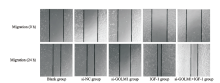
Fig. 3
Comparison of the migration ability of lung adenocarcinoma A549 cells in each group"
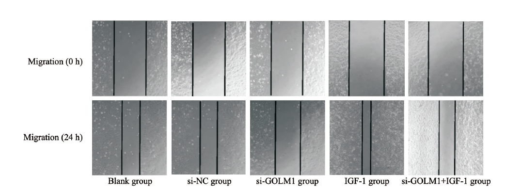
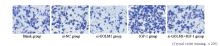
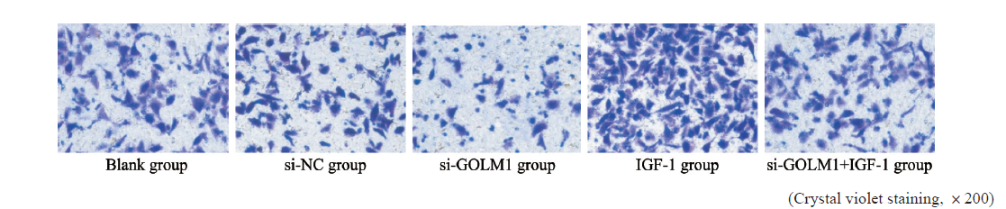
Fig. 4
Comparison of the invasion ability of lung adenocarcinoma A549 cells in each group"
Tab. 4
Comparison of scratch healing rate and number of invasive cells in lung adenocarcinoma A549 cells in each group ($\bar{x}±s$, n=6)"
| Group | Scratch healing rate/% | Number of invasion cells |
|---|---|---|
| Blank group | 46.25±3.08 | 65.86±4.28 |
| si-NC group | 44.73±3.23 | 68.34±3.98 |
| si-GOLM1 group | 18.65±1.79ab | 29.06±2.55ab |
| IGF-1 group | 79.62±5.62abc | 112.39±7.87abc |
| si-GOLM1+IGF-1 group | 52.27±3.35c | 75.66±4.43c |
| F value | 215.315 | 216.280 |
| P value | <0.001 | <0.001 |
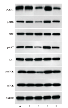
Fig. 5
Western blot method was used to detect GOLM1, p-PI3K, PI3K, p-AKT, AKT, p-mTOR and mTOR protein expression in lung adenocarcinoma A549 cells in each group A: Blank group; B: si-NC group; C: si-GOLM1 group; D: IGF-1 group; E: si-GOLM1+IGF-1 group."
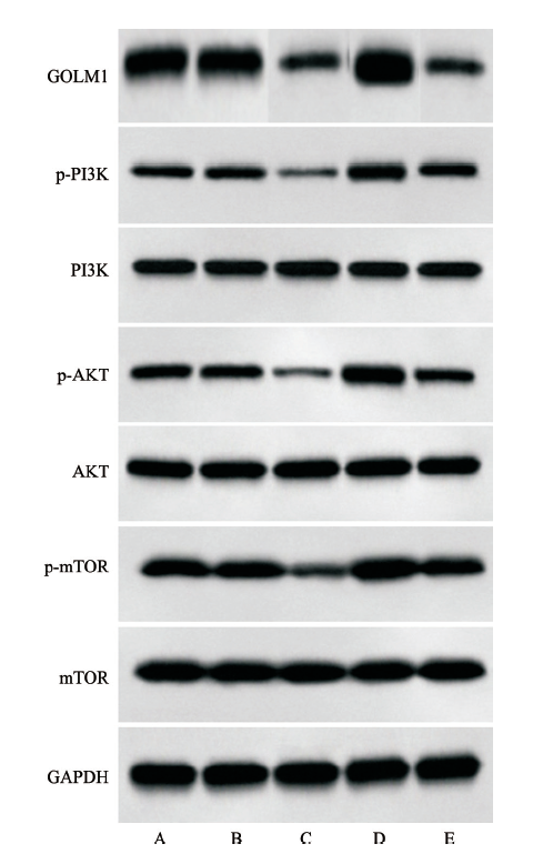
Tab. 5
Comparison of GOLM1 and PI3K/AKT/mTOR signaling pathway related protein levels in lung adenocarcinoma A549 cells in each group ($\bar{x}±s$, n=6)"
| Group | GOLM1/GAPDH | p-PI3K/PI3K | p-AKT/AKT | p-mTOR/mTOR |
|---|---|---|---|---|
| Blank group | 1.51±0.12 | 0.45±0.03 | 0.68±0.04 | 0.79±0.05 |
| si-NC group | 1.52±0.14 | 0.48±0.02 | 0.71±0.05 | 0.78±0.06 |
| si-GOLM1 group | 0.63±0.04ab | 0.15±0.01ab | 0.18±0.01ab | 0.25±0.03ab |
| IGF-1 group | 1.56±0.11c | 0.83±0.05abc | 1.08±0.06abc | 1.21±0.11abc |
| si-GOLM1+IGF-1 group | 0.65±0.05ab | 0.51±0.02c | 0.72±0.05c | 0.83±0.06c |
| F value | 142.249 | 406.605 | 300.233 | 154.652 |
| P value | <0.001 | <0.001 | <0.001 | <0.001 |
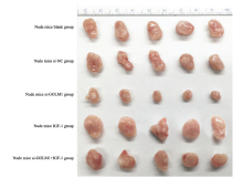
Fig. 6
Comparison of the growth of xenagraft tumors in BALB/c nude mice in each group"
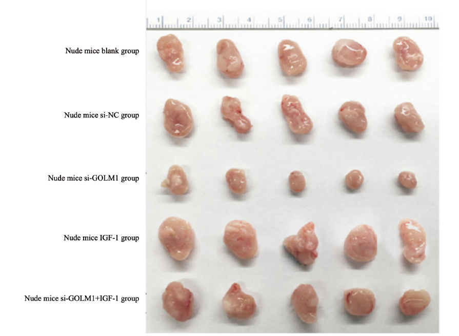
Tab. 6
Comparison of the mass and volume of transplanted tumors in BALB/c nude mice in each group ($\bar{x}±s$, n=6)"
| Group | Tumor quality m/g | Tumor volume V/mm3 |
|---|---|---|
| Nude mice blank group | 0.86±0.04 | 175.51±8.16 |
| Nude mice si-NC group | 0.89±0.05 | 181.63±10.20 |
| Nude mice si-GOLM1 group | 0.35±0.01ab | 71.43±2.04ab |
| Nude mice IGF-1 group | 1.23±0.11abc | 251.02±22.45abc |
| Nude mice si-GOLM1+IGF-1 group | 0.95±0.07c | 193.88±12.14c |
| F value | 143.604 | 153.473 |
| P value | <0.001 | <0.001 |
| [1] |
LUO C, LEI M, ZHANG Y, et al. Systematic construction and validation of an immune prognostic model for lung adenocarcinoma[J]. J Cell Mol Med, 2020, 24(2): 1233-1244.
doi: 10.1111/jcmm.v24.2 |
| [2] |
KLECZKO E K, KWAK J W, SCHENK E L, et al. Targeting the complement pathway as a therapeutic strategy in lung cancer[J]. Front Immunol, 2019, 10: 954.
doi: 10.3389/fimmu.2019.00954 |
| [3] | YAN J L, ZHOU B H, GUO L, et al. GOLM1 upregulates expression of PD-L1 through EGFR/STAT3 pathway in hepatocellular carcinoma[J]. Am J Cancer Res, 2020, 10(11): 3705-3720. |
| [4] | 郑兴, 吴兆红, 陈岗东, 等. miRNA27a靶向GOLM1对非小细胞肺癌生物行为学的影响[J]. 中国免疫学杂志, 2020, 36(1): 52-56. |
| ZHENG X, WU Z H, CHEN G D, et al. Effect of miRNA27a targeting GOLM1 on biobehavioral of non-small cell lung cancer[J]. Chin J Immunol, 2020, 36(1): 52-56. | |
| [5] | 曾建昌, 刘艳, 杨俊, 等. 复方苦参注射液联合奥希替尼介导PI3K/AKT/mTOR信号通路在肺腺癌细胞H1975中的作用机制[J]. 中国免疫学杂志, 2021, 37(10): 1191-1195. |
| ZENG J C, LIU Y, YANG J, et al. Mechanism of compound sophora flavescens injection combined with osimertinib in lung adenocarcinoma cell H1975 via PI3K/AKT/mTOR signaling pathway[J]. Chin J Immunol, 2021, 37(10): 1191-1195. | |
| [6] | 王永贵. SLC25A22在骨肉瘤组织中的表达及其与预后的相关性[J]. 现代肿瘤医学, 2021, 29(17): 3093-3096. |
| WANG Y G. Expression of SLC25A22 in osteosarcoma and its correlation with prognosis[J]. J Mod Oncol, 2021, 29(17): 3093-3096. | |
| [7] |
RONG L, LI Z D, LENG X, et al. Salidroside induces apoptosis and protective autophagy in human gastric cancer AGS cells through the PI3K/AKT/mTOR pathway[J]. Biomed Pharmacother, 2020, 122: 109726.
doi: 10.1016/j.biopha.2019.109726 |
| [8] |
WANG Y Q, HUANG J, WU Q, et al. Downregulation of breast cancer resistance protein by long-term fractionated radiotherapy sensitizes lung adenocarcinoma to SN-38[J]. Invest New Drugs, 2021, 39(2): 458-468.
doi: 10.1007/s10637-020-01003-3 |
| [9] |
ZHANG R, ZHU Z, SHEN W Z, et al. Golgi membrane protein 1 (GOLM1) promotes growth and metastasis of breast cancer cells via regulating matrix metalloproteinase-13 (MMP13)[J]. Med Sci Monit, 2019, 25: 847-855.
doi: 10.12659/MSM.911667 |
| [10] |
YE Q H, ZHU W W, ZHANG J B, et al. GOLM1 modulates EGFR/RTK cell-surface recycling to drive hepatocellular carcinoma metastasis[J]. Cancer Cell, 2016, 30(3): 444-458.
doi: 10.1016/j.ccell.2016.07.017 |
| [11] | 翟维佳, 封全灵, 张慧芳, 等. GOLM1对宫颈癌上皮间质转化的影响及其与临床病理特征的关系[J]. 肿瘤学杂志, 2019, 25(11): 976-979. |
| ZHAI W J, FENG Q L, ZHANG H F, et al. Effect of GOLM1 expression on epithelial-mesenchymal transition and its relationship with clinicopathological features of cervical cancer[J]. J Chin Oncol, 2019, 25(11): 976-979. | |
| [12] | 余展鹏, 江雁琼. 基于数据挖掘分析GOLM1基因在前列腺癌中的表达及其临床意义[J]. 中国癌症防治杂志, 2019, 11(2): 158-162. |
| YU Z P, JIANG Y Q. Analysis of GOLM1 gene expression in prostate cancer and its effect on prognosis based on data mining[J]. Chin J Oncol Prev Treat, 2019, 11(2): 158-162. | |
| [13] | ARUNA, LI L M. Overexpression of Golgi membrane protein 1 promotes non-small-cell carcinoma aggressiveness by regulating the matrix metallopeptidase 13[J]. Am J Cancer Res, 2018, 8(3): 551-565. |
| [14] | ZHANG S K, GE W M, ZOU G Y, et al. MiR-382 targets GOLM1 to inhibit metastasis of hepatocellular carcinoma and its down-regulation predicts a poor survival[J]. Am J Cancer Res, 2018, 8(1): 120-131. |
| [15] | YANG L Q, LUO P C, SONG Q, et al. DNMT1/miR-200a/GOLM1 signaling pathway regulates lung adenocarcinoma cells proliferation[J]. Biomedecine Pharmacother, 2018, 99: 839-847. |
| [16] |
CIRONE M. Cancer cells dysregulate PI3K/AKT/mTOR pathway activation to ensure their survival and proliferation: mimicking them is a smart strategy of gammaherpesviruses[J]. Crit Rev Biochem Mol Biol, 2021, 56(5): 500-509.
doi: 10.1080/10409238.2021.1934811 |
| [17] |
GU Z H, YOU Z X, YANG Y C, et al. Inhibition of MicroRNA miR-101-3p on prostate cancer progression by regulating cullin 4B (CUL4B) and PI3K/AKT/mTOR signaling pathways[J]. Bioengineered, 2021, 12(1): 4719-4735.
doi: 10.1080/21655979.2021.1949513 |
| [18] | DENG L, WU X R, ZHU X J, et al. Combination effect of curcumin with docetaxel on the PI3K/AKT/mTOR pathway to induce autophagy and apoptosis in esophageal squamous cell carcinoma[J]. Am J Transl Res, 2021, 13(1): 57-72. |
| [19] | 炎士珂, 冯婧文, 昌娇, 等. FGFC1经PI3K/AKT/mTOR途径抑制非小细胞肺癌细胞增殖和迁移[J]. 中国生物化学与分子生物学报, 2021, 37(8): 1069-1077. |
| YAN S K, FENG J W, CHANG J, et al. FGFC1 inhibits proliferation and migration of non-small cell lung cancer cells via the PI3K/AKT/mTOR signaling pathway[J]. Chin J Biochem Mol Biol, 2021, 37(8): 1069-1077. | |
| [20] | 孟东雪, 郭玉荣, 罗斌军, 等. 扶正抑瘤汤通过PI3K/AKT/mTOR信号通路抑制非小细胞肺癌增殖、凋亡及自噬[J]. 中国癌症防治杂志, 2021, 13(2): 177-182. |
| MENG D X, GUO Y R, LUO B J, et al. Fuzheng Yiliu decoction inhibits the proliferation, apoptosis and autophagy of non-small cell lung cancer through PI3K/AKT/mTOR signaling pathway[J]. Chin J Oncol Prev Treat, 2021, 13(2): 177-182. | |
| [21] | 丁志丹, 方泽民, 王旭广, 等. 贝母素乙调控PI3K/AKT/mTOR通路减缓上皮-间质转化进程抑制人肺癌A549细胞侵袭及迁移的研究[J]. 中草药, 2019, 50(6): 1382-1387. |
| DING Z D, FANG Z M, WANG X G, et al. Inhibition of peiminine on invasion and migration of human lung cancer A549 cells by decreasing epithelial-mesenchymal transition process via PI3K/AKT/mTOR pathway[J]. Chin Tradit Herb Drugs, 2019, 50(6): 1382-1387. |
| [1] | SUN Rongqi, SONG Ning, ZHENG Wentian, ZHANG Xinyue, LI Minmin, GONG Hui, JIANG Yingying. Effect of long noncoding RNA FLJ30679 on proliferation and migration of oral squamous cell carcinoma cells [J]. China Oncology, 2024, 34(5): 439-450. |
| [2] | XIONG Jiayan, LEI Wei, YOU Bo, ZHANG Zhenxin, XIE Haijing, SHAN Ying, XIA Tian, ZHOU Yong. Study on the mechanism of DDX6 promoting proliferation and migration of nasopharyngeal carcinoma cells by regulating stability of CKMT1A mRNA [J]. China Oncology, 2024, 34(5): 451-459. |
| [3] | ZHOU Xueqin, LUAN Yanchao, ZHAO Li, RONG Chaochao, YANG Na. Expression of CDC20 in lung adenocarcinoma tissues and its effect on the proliferation and invasion of lung adenocarcinoma cells [J]. China Oncology, 2024, 34(5): 460-472. |
| [4] | JIA Liqing, GE Xiaolu, JIANG Lin, ZHOU Xiaoyan. Effects of lncRNA PKD2-2-3 on cell proliferation, clone formation, migration, and invasion of lung adenocarcinoma [J]. China Oncology, 2023, 33(8): 717-725. |
| [5] | DONG Hao, QIU Yonggang, WANG Xinbin, YANG Junjie, LOU Cuncheng, YIN Lekang, YE Xiaodan. Predictive value of logistic regression model based on high-resolution CT signs for high-grade pattern in stage ⅠA lung adenocarcinoma [J]. China Oncology, 2023, 33(8): 768-775. |
| [6] | LIU Xiaoli, CHAI Wenjun, SUN Lei, YAN Mingxia, PAN Hongyu, SUN Yuexi. Analysis of differential splicing gene by regulation of splicing regulatory protein KHSRP in lung adenocarcinoma [J]. China Oncology, 2023, 33(7): 637-645. |
| [7] | CHEN Hong, CHEN Junxia. Effect of hsa_circ_0001573 on biological behaviors of breast cancer cells and its molecular mechanism [J]. China Oncology, 2023, 33(4): 342-353. |
| [8] | MU Jiaqian, TENG Xiaoyan, WEI Lirong, QIU Rong, GUI Pengcheng, DU Yuzhen. The role and application value of integrin β3 in bone metastasis of lung adenocarcinoma [J]. China Oncology, 2022, 32(4): 351-356. |
| [9] | DUAN Yuqing, XIA Ning, JIA Yunlong, ZHENG Wenya, LIU Lihua. SRSF1 promotes proliferation, invasion and migration of esophageal squamous cell carcinoma Eca9706 cells by regulating VEGFA mRNA alternative splicing [J]. China Oncology, 2022, 32(3): 191-199. |
| [10] | HOU Qinghua, ZHONG Yanfeng, LIU Linzhuang, WU Liusheng, LIU Jixian. Expression, prognostic value of CBX3 in lung adenocarcinoma and its effect on biological behavior of cancer cells [J]. China Oncology, 2022, 32(2): 152-160. |
| [11] | ZHANG Longfu, LIU Jie, NI Zheng, LU Xinyuan, HU Bin, WANG Hao, FENG Mingxiang, ZHANG Yong. Development and validation of a nomogram for predicting spread through air spaces in stage ⅠA lung adenocarcinoma [J]. China Oncology, 2022, 32(12): 1210-1217. |
| [12] | LI Haizhou , ZHANG Yanwei , XU Yingjie , YANG Men , ZHANG Lei , HAN Jingjun . miR-933 inhibits proliferation, migration and invasion of lung cancer cell lines by regulation of KLF6 gene [J]. China Oncology, 2021, 31(7): 581-588. |
| [13] | XIE Jinfang , CAO Chunyu , REN Xue , TIAN Jiajun , LÜ Yafeng , HUANG Xiaofei . Effects of sulforaphane on epithelial-mesenchymal transition, proliferation and migration of mouse breast cancer 4T1 cells [J]. China Oncology, 2021, 31(7): 605-615. |
| [14] | HU Guannan , CHEN Lei , ZHOU Liangping , ZHOU Zhengrong . A case report of pulmonary malignant melanoma complicated with lung adenocarcinoma and literature review [J]. China Oncology, 2021, 31(7): 647-650. |
| [15] | HU Yaqiong , BAI Jun , CHEN Lin , CHEN Xinlu , ZHANG Liping , ZHOU Dandan , WANG Yu , YIN Chonggao , LI Hongli , LIU Yuqing . miR-625-5p promotes proliferation and invasion of lung adenocarcinoma by targeting PRKACA [J]. China Oncology, 2021, 31(6): 447-454. |
| Viewed | ||||||
|
Full text |
|
|||||
|
Abstract |
|
|||||
沪ICP备12009617
Powered by Beijing Magtech Co. Ltd