Welcome to China Oncology,
China Oncology ›› 2023, Vol. 33 ›› Issue (8): 768-775.doi: 10.19401/j.cnki.1007-3639.2023.08.005
• Article • Previous Articles Next Articles
DONG Hao1( ), QIU Yonggang1, WANG Xinbin1, YANG Junjie2, LOU Cuncheng1, YIN Lekang3(
 ), YE Xiaodan3,4,5()
Received:2023-02-03
Revised:2023-05-12
Online:2023-08-30
Published:2023-09-01
Contact:
YIN Lekang, YE Xiaodan
E-mail:yuanyxd@163.com
Share article
CLC Number:
DONG Hao, QIU Yonggang, WANG Xinbin, YANG Junjie, LOU Cuncheng, YIN Lekang, YE Xiaodan. Predictive value of logistic regression model based on high-resolution CT signs for high-grade pattern in stage ⅠA lung adenocarcinoma[J]. China Oncology, 2023, 33(8): 768-775.
Tab. 1
Clinical and pathological characteristics of patients"
| Item | Total | HGP (n=88) | n-HGP (n=357) | P value |
|---|---|---|---|---|
| Age/year | 58±11 | 57±12 | 61±9 | 0.037△ |
| Gender | 0.015# | |||
| Male | 158 | 41 | 117 | |
| Female | 287 | 47 | 240 | |
| Smoking history | 0.003# | |||
| No | 390 | 69 | 321 | |
| Yes | 55 | 19 | 36 | |
| Location | 0.401# | |||
| Left upper lobe | 108 | 22 | 86 | |
| Left lower lobe | 69 | 17 | 52 | |
| Right upper lobe | 148 | 29 | 119 | |
| Right middle lobe | 36 | 9 | 27 | |
| Right lower lobe | 84 | 11 | 73 | |
| Clinical T staging | <0.001# | |||
| T1a | 121 | 10 | 111 | |
| T1b | 258 | 54 | 204 | |
| T1c | 66 | 24 | 42 | |
| Pure or mix pattern | 0.008* | |||
| Pure | 27 | 0 | 27 | |
| Mix | 418 | 88 | 330 | |
| Major histological patterns | <0.001* | |||
| Lepidic predominant | 52 | 1 | 51 | |
| Acinar predominant | 348 | 71 | 280 | |
| Papillary predominant | 32 | 6 | 26 | |
| Micropapillary | 4 | 4 | 0 | |
| Solid predominant | 9 | 9 | 0 |
Tab. 2
Observer consistency test results for CT features"
| Item | Case n | SE | Kappa value | 95% CI |
|---|---|---|---|---|
| Density | 443 | 0.012 | 0.983 | 0.955-1.000 |
| Shape | 440 | 0.013 | 0.972 | 0.942-0.994 |
| Lobulation | 422 | 0.022 | 0.892 | 0.847-0.933 |
| Spiculation | 430 | 0.018 | 0.924 | 0.886-0.956 |
| Vacuole | 427 | 0.048 | 0.791 | 0.685-0.837 |
| Air bronchogram | 435 | 0.015 | 0.955 | 0.926-0.982 |
| Pleural indentation | 442 | 0.008 | 0.986 | 0.968-1.000 |
Tab. 3
Comparison of CT features of lesions between the HGP and n-HGP groups"
| Item | Total | HGP (n=88) | n-HGP (n=357) | P value |
|---|---|---|---|---|
| Tumor size D/mm | 14±5 | 17±5 | 14±5 | <0.001△ |
| Density | <0.001# | |||
| SSN | 377 | 45 | 332 | |
| SN | 68 | 43 | 25 | |
| Shape | 0.003# | |||
| Round or oval | 129 | 14 | 115 | |
| Irregular | 316 | 74 | 242 | |
| Lobulation | <0.001# | |||
| No | 287 | 25 | 262 | |
| Yes | 158 | 63 | 95 | |
| Spiculation | <0.001# | |||
| No | 296 | 32 | 264 | |
| Yes | 149 | 56 | 93 | |
| Vacuole | 0.764# | |||
| No | 406 | 81 | 325 | |
| Yes | 39 | 7 | 32 | |
| Air bronchogram | 0.162# | |||
| No | 242 | 42 | 200 | |
| Yes | 203 | 46 | 157 | |
| Pleural indentation | <0.001# | |||
| No | 256 | 33 | 223 | |
| Yes | 189 | 55 | 134 |

Fig. 1
CT and pathology images of SSN in the upper lobe of the left lung A 55-year-old male patient, no smoking history. A: HRCT transection lung window showed SSN in the upper lobe of the left lung, with a length diameter of about 15 mm and no lobes around it; B: Reconstructed image (coronal position), nodules did not show lobulation signs; C: Pathological diagram, the result was acinar predominant invasive adenocarcinoma (H-E, ×100)."
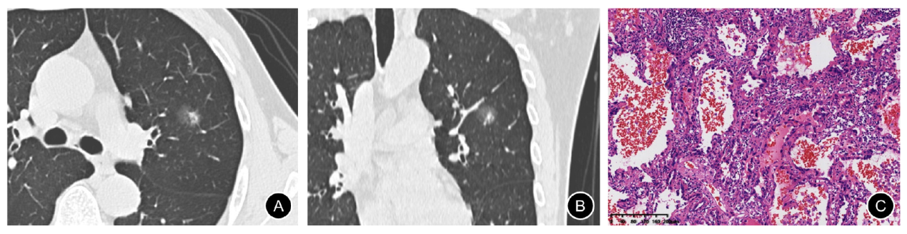
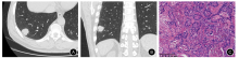
Fig. 2
CT and pathology images of SN in the lower lobe of the right lung A 58-year-old male patient with a smoking history of 30 years. A: HRCT transection lung window showed SN in the upper lobe of the left lung, with a long diameter of about 26 mm, and the lobulation sign was visible around the periphery; B: Reconstruction image (coronal position), nodules showed lobulation signs; C: Pathological diagram, the result was solid predominant invasive adenocarcinoma (H-E, ×100)."
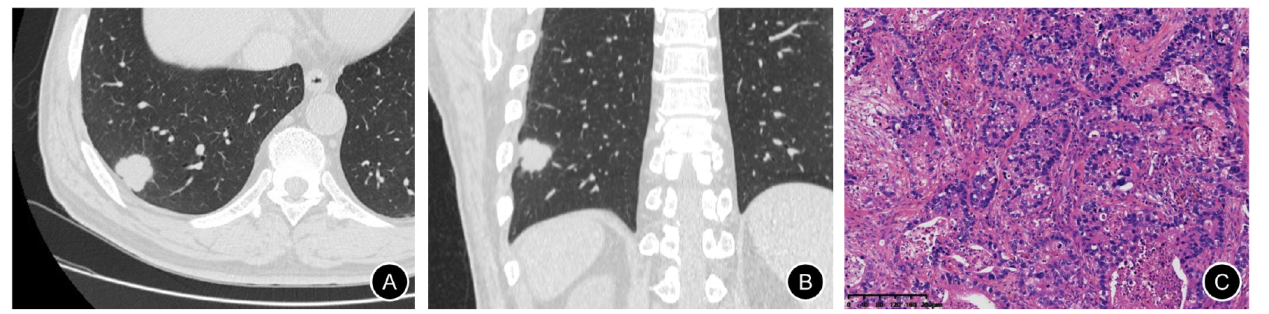

Fig. 3
ROC curves predicted by clinical model, CT model and clinical-CT model"
Tab. 4
Comparison of diagnostic performance of CT model and univariate signs in predicting HGP"
| Item | AUC (95% CI) | Sensitivity/% | Specificity/% |
|---|---|---|---|
| CT model | 0.838 (0.788-0.889) | 70.5 | 84.9 |
| Tumor size | 0.695 (0.636-0.755) | 75.0 | 56.4 |
| Density | 0.709 (0.640-0.778) | 48.9 | 97.0 |
| Shape | 0.582 (0.519-0.644) | 84.1 | 32.2 |
| Lobulation | 0.725 (0.664-0.785) | 71.6 | 73.4 |
| Spiculation | 0.688 (0.624-0.752) | 63.6 | 73.9 |
| Pleural indentation | 0.625 (0.560-0.690) | 62.5 | 62.5 |
| [1] |
TRAVIS W D, BRAMBILLA E, NOGUCHI M, et al. International Association for the Study of Lung Cancer/American Thoracic Society/European Respiratory Society international multidisciplinary classification of lung adenocarcinoma[J]. J Thorac Oncol, 2011, 6(2): 244-285.
doi: 10.1097/JTO.0b013e318206a221 pmid: 21252716 |
| [2] |
TAKAHASHI Y, EGUCHI T, KAMEDA K, et al. Histologic subtyping in pathologic stage Ⅰ-ⅡA lung adenocarcinoma provides risk-based stratification for surveillance[J]. Oncotarget, 2018, 9(87): 35742-35751.
doi: 10.18632/oncotarget.v9i87 |
| [3] |
YASUKAWA M, OHBAYASHI C, KAWAGUCHI T, et al. Analysis of histological grade in resected lung-invasive adenocarcinoma[J]. Anticancer Res, 2019, 39(3): 1491-1500.
doi: 10.21873/anticanres.13267 pmid: 30842187 |
| [4] |
JEON H W, KIM Y D, SIM S B, et al. Significant difference in recurrence according to the proportion of high grade patterns in stage ⅠA lung adenocarcinoma[J]. Thorac Cancer, 2021, 12(13): 1952-1958.
doi: 10.1111/tca.v12.13 |
| [5] |
VAN SCHIL P E, ASAMURA H, RUSCH V W, et al. Surgical implications of the new IASLC/ATS/ERS adenocarcinoma classification[J]. Eur Respir J, 2012, 39(2): 478-486.
doi: 10.1183/09031936.00027511 pmid: 21828029 |
| [6] |
HUNG J J, JENG W J, CHOU T Y, et al. Prognostic value of the new International Association for the Study of Lung Cancer/American Thoracic Society/European Respiratory Society lung adenocarcinoma classification on death and recurrence in completely resected stage Ⅰ lung adenocarcinoma[J]. Ann Surg, 2013, 258(6): 1079-1086.
doi: 10.1097/SLA.0b013e31828920c0 |
| [7] | HUNG J J, YEH Y C, JENG W J, et al. Predictive value of the International Association for the Study of Lung Cancer/American Thoracic Society/European Respiratory Society classification of lung adenocarcinoma in tumor recurrence and patient survival[J]. J Clin Oncol, 2014, 32(22): 2357-2364. |
| [8] |
CHOI Y, KIM J, PARK H, et al. Rethinking a non-predominant pattern in invasive lung adenocarcinoma: prognostic dissection focusing on a high-grade pattern[J]. Cancers (Basel), 2021, 13(11): 2785.
doi: 10.3390/cancers13112785 |
| [9] |
TAKAHASHI Y, KURODA H, OYA Y, et al. Challenges for real-time intraoperative diagnosis of high risk histology in lung adenocarcinoma: a necessity for sublobar resection[J]. Thorac Cancer, 2019, 10(8): 1663-1668.
doi: 10.1111/1759-7714.13133 pmid: 31287246 |
| [10] |
ANDRE L, MOREIRA, MD P, et al. A grading system for invasive pulmonary adenocarcinoma: a proposal from the international association for the study of lung cancer pathology committee[J]. J Thorac Oncol, 2020, 15(10): 1599-1610.
doi: S1556-0864(20)30468-8 pmid: 32562873 |
| [11] |
SUNG H, FERLAY J, SIEGEL R L, et al. Global cancer statistics 2020: GLOBOCAN estimates of incidence and mortality worldwide for 36 cancers in 185 countries[J]. CA Cancer J Clin, 2021, 71(3): 209-249.
doi: 10.3322/caac.v71.3 |
| [12] |
KUHN E, MORBINI P, CANCELLIERI A, et al. Adenocarcinoma classification: patterns and prognosis[J]. Pathologica, 2018, 110(1): 5-11.
pmid: 30259909 |
| [13] |
ZHANG Y, QIANG J W, SHEN Y, et al. Using air bronchograms on multi-detector CT to predict the invasiveness of small lung adenocarcinoma[J]. Eur J Radiol, 2016, 85(3): 571-577.
doi: 10.1016/j.ejrad.2015.12.014 pmid: 26860669 |
| [14] | 金鑫, 赵绍宏, 高洁, 等. 纯磨玻璃密度肺腺癌病理分类及影像表现特点分析[J]. 中华放射学杂志, 2014, 48(4): 283-287 |
| JIN X, ZHAO S H, GAO J, et al. Pathological classification and imaging characteristics of early-stage lung adenocarcinoma with pure ground-glass opacity[J]. Chin J Radiol, 2014, 48(4): 283-287 | |
| [15] | 蔡蒙婷, 纪晓微, 傅钢泽, 等. 含气腔型浸润性肺小腺癌的CT表现与病理分型的对照研究[J]. 中华放射学杂志, 2019, 53(10): 886-891. |
| [16] |
ZHANG D G, CHEN X C, ZHU D X, et al. Intrapulmonary lymph node metastasis is common in clinically staged ⅠA adenocarcinoma of the lung[J]. Thorac Cancer, 2019, 10(2): 123-127.
doi: 10.1111/tca.2019.10.issue-2 |
| [17] |
YU Y, JIAN H, SHEN L, et al. Lymph node involvement influenced by lung adenocarcinoma subtypes in tumor size ≤3 cm disease: a study of 2 268 cases[J]. Eur J Surg Oncol, 2016, 42(11): 1714-1719.
doi: 10.1016/j.ejso.2016.02.247 |
| [18] |
MIKUBO M, NAITO M, MATSUI Y, et al. Relevance of intraoperative pleural lavage cytology and histologic subtype in lung adenocarcinoma[J]. Ann Thorac Surg, 2018, 106(6): 1654-1660.
doi: S0003-4975(18)31256-6 pmid: 30227125 |
| [19] |
LEE G, CHOI E R, LEE H Y, et al. Pathologic heterogeneity of lung adenocarcinomas: a novel pathologic index predicts survival[J]. Oncotarget, 2016, 7(43): 70353-70363.
doi: 10.18632/oncotarget.11857 pmid: 27612421 |
| [20] |
SUNG Y E, LEE K Y, MOON Y. The prognostic utility of the histologic subtype of stage Ⅰ lung adenocarcinoma may be diminished when using only the invasive component to determine tumor size for tumor node metastasis (TNM) staging[J]. J Thorac Dis, 2021, 13(5): 2910-2922.
doi: 10.21037/jtd |
| [21] |
CHEN L W, YANG S M, WANG H J, et al. Prediction of micropapillary and solid pattern in lung adenocarcinoma using radiomic values extracted from near-pure histopathological subtypes[J]. Eur Radiol, 2021, 31(7): 5127-5138.
doi: 10.1007/s00330-020-07570-6 |
| [22] |
SUN F H, HUANG Y W, YANG X D, et al. Solid component ratio influences prognosis of GGO-featured ⅠA stage invasive lung adenocarcinoma[J]. Cancer Imaging, 2020, 20(1): 87.
doi: 10.1186/s40644-020-00363-6 |
| [23] |
MIYOSHI T, AOKAGE K, KATSUMATA S, et al. Ground-glass opacity is a strong prognosticator for pathologic stage ⅠA lung adenocarcinoma[J]. Ann Thorac Surg, 2019, 108(1): 249-255.
doi: 10.1016/j.athoracsur.2019.01.079 |
| [24] |
MA X, ZHOU S, HUANG L, et al. Assessment of relationships among clinicopathological characteristics, morphological computer tomography features, and tumor cell proliferation in stage Ⅰ lung adenocarcinoma[J]. J Thorac Dis, 2021, 13(5): 2844-2857.
doi: 10.21037/jtd |
| [1] | WENG Junyong, YE Zilan, ZHANG Ruoxin, LIU Qi, LI Xinxiang. Exploring the guiding role of the number of adverse pathological features in risk stratification for recurrence of stage Ⅰ-Ⅲ colorectal cancer: a retrospective cohort study of 9 875 cases [J]. China Oncology, 2024, 34(6): 527-536. |
| [2] | ZHANG Ruoxin, YE Zilan, WENG Junyong, LI Xinxiang. Correlation study between advanced age and inferior prognosis in stage Ⅱ colorectal cancer patients [J]. China Oncology, 2024, 34(5): 485-492. |
| [3] | OUYANG Fei, WANG Yang, CHEN Yu, PEI Guoqing, WANG Ling, ZHANG Yang, SHI Lei. Construction of the prediction model of breast cancer bone metastasis based on machine learning [J]. China Oncology, 2024, 34(10): 903-914. |
| [4] | JIA Liqing, GE Xiaolu, JIANG Lin, ZHOU Xiaoyan. Effects of lncRNA PKD2-2-3 on cell proliferation, clone formation, migration, and invasion of lung adenocarcinoma [J]. China Oncology, 2023, 33(8): 717-725. |
| [5] | LIU Xiaoli, CHAI Wenjun, SUN Lei, YAN Mingxia, PAN Hongyu, SUN Yuexi. Analysis of differential splicing gene by regulation of splicing regulatory protein KHSRP in lung adenocarcinoma [J]. China Oncology, 2023, 33(7): 637-645. |
| [6] | YANG Wenxiao, GUO Linwei, LING Hong, HU Xin. Characterization of immune microenvironment identifies prognostic and immunotherapy benefit for trastuzumab-based therapy [J]. China Oncology, 2023, 33(5): 484-498. |
| [7] | CHEN Yingyao, CHU Xiangling, YU Xin, SU Chunxia. Advances in models predicting efficacy of immune checkpoint inhibitors [J]. China Oncology, 2023, 33(1): 61-70. |
| [8] | XU Yuchen, CHENG Leilei, WANG Yan, LIN Jinyi, CHEN Jiahui, CHEN Yifan, ZHOU Yuhong, LIU Tianshu, GE Junbo. Predictive value of sST2 level in immune-related adverse events [J]. China Oncology, 2022, 32(8): 712-718. |
| [9] | MU Jiaqian, TENG Xiaoyan, WEI Lirong, QIU Rong, GUI Pengcheng, DU Yuzhen. The role and application value of integrin β3 in bone metastasis of lung adenocarcinoma [J]. China Oncology, 2022, 32(4): 351-356. |
| [10] | ZHU Haipeng, HU Jun, JIANG Min, CAI Ruonan, WANG Junqiao, LI Li. A study on mechanism of GOLM1 regulating PI3K/AKT/mTOR signaling pathway to promote proliferation, invasion and migration of lung adenocarcinoma cells [J]. China Oncology, 2022, 32(3): 207-217. |
| [11] | HOU Qinghua, ZHONG Yanfeng, LIU Linzhuang, WU Liusheng, LIU Jixian. Expression, prognostic value of CBX3 in lung adenocarcinoma and its effect on biological behavior of cancer cells [J]. China Oncology, 2022, 32(2): 152-160. |
| [12] | ZHANG Longfu, LIU Jie, NI Zheng, LU Xinyuan, HU Bin, WANG Hao, FENG Mingxiang, ZHANG Yong. Development and validation of a nomogram for predicting spread through air spaces in stage ⅠA lung adenocarcinoma [J]. China Oncology, 2022, 32(12): 1210-1217. |
| [13] | HU Guannan , CHEN Lei , ZHOU Liangping , ZHOU Zhengrong . A case report of pulmonary malignant melanoma complicated with lung adenocarcinoma and literature review [J]. China Oncology, 2021, 31(7): 647-650. |
| [14] | HU Yaqiong , BAI Jun , CHEN Lin , CHEN Xinlu , ZHANG Liping , ZHOU Dandan , WANG Yu , YIN Chonggao , LI Hongli , LIU Yuqing . miR-625-5p promotes proliferation and invasion of lung adenocarcinoma by targeting PRKACA [J]. China Oncology, 2021, 31(6): 447-454. |
| [15] | PENG Zishan, KONG Hui, BAO Zhen, ZHAO Hongxing, LIU Xin, LU Shaohua. Clinicopathological analysis of 83 cases of multifocal lung adenocarcinoma [J]. China Oncology, 2021, 31(5): 408-418. |
| Viewed | ||||||
|
Full text |
|
|||||
|
Abstract |
|
|||||
沪ICP备12009617
Powered by Beijing Magtech Co. Ltd