Welcome to China Oncology,
China Oncology ›› 2024, Vol. 34 ›› Issue (9): 857-872.doi: 10.19401/j.cnki.1007-3639.2024.09.006
• Article • Previous Articles Next Articles
HUANG Haozhe1( ), CHEN Hong2(), ZHENG Dezhong3, CHEN Chao1, WANG Ying1, XU Lichao1, WANG Yaohui1, HE Xinhong1, YANG Yuanyuan3, LI Wentao1()
Received:2024-05-07
Revised:2024-06-13
Online:2024-09-30
Published:2024-10-11
Contact:
LI Wentao
Share article
CLC Number:
HUANG Haozhe, CHEN Hong, ZHENG Dezhong, CHEN Chao, WANG Ying, XU Lichao, WANG Yaohui, HE Xinhong, YANG Yuanyuan, LI Wentao. A CT-based radiomics nomogram for predicting local tumor progression of colorectal cancer lung metastases treated with radiofrequency ablation[J]. China Oncology, 2024, 34(9): 857-872.

Fig. 1
Workflow of the study"
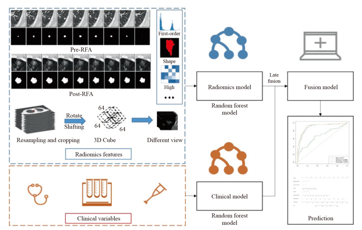
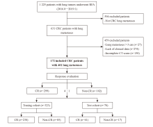
Fig. 2
Flow diagram of the enrolment patients"
Tab. 1
Characteristics of CRC patients with lung metastases"
| Characteristic | Training dataset (N=323) | Test dataset (N=78) | P value | Total (N=401) |
|---|---|---|---|---|
| Pre-RFA clinical characteristics | ||||
| Gender n | 0.947 | |||
| Male | 185 | 45 | 230 | |
| Female | 138 | 33 | 171 | |
| Age/year M (Q1, Q3) | 57 (50, 65) | 61 (52, 66.25) | 0.120 | |
| Initial tumor location n | 0.166 | |||
| Rectum | 224 | 44 | 268 | |
| Sigmoid-left colon | 40 | 15 | 55 | |
| Transverse-right colon | 57 | 18 | 75 | |
| Caecum | 2 | 1 | 3 | |
| Tumor biomarkers M (Q1, Q3) | ||||
| CEA/(ng·mL-1) | 4.39 (2.07, 13.33) | 3.96 (2.08, 11.38) | 0.628 | |
| CA 19-9/(U·mL-1) | 10.71 (7.58, 19.50) | 12.60 (6.83, 23.25) | 0.928 | |
| Number of recurrence n | 0.351 | |||
| At 1 year | 75 | 17 | 92 | |
| At 2 years | 82 | 17 | 99 | |
| At 3 years | 85 | 17 | 102 | |
| Follow-time/month M (95% CI) | 25 (22.49, 27.51) | 19 (18.43, 19.57) | ||
| Pre-RFA characteristics of lung metastases | ||||
| Nodule size/mm n | 0.064 | |||
| <10 | 156 | 35 | 191 | |
| 10-19 | 118 | 31 | 149 | |
| 20-30 | 49 | 12 | 61 | |
| Location n | 0.183 | |||
| RUL | 76 | 15 | 91 | |
| RML | 36 | 8 | 44 | |
| RLL | 63 | 10 | 73 | |
| LUL | 77 | 18 | 95 | |
| LLL | 71 | 27 | 98 | |
| Distance 1/cm n | 0.454 | |||
| ≥1 | 266 | 67 | 333 | |
| <1 | 57 | 11 | 68 | |
| Distance 2/cm n | 0.399 | |||
| ≥1 | 124 | 34 | 158 | |
| <1 | 199 | 44 | 243 | |
| Immediate post-RFA characteristics | ||||
| Immediate pneumothorax n | 0.851 | |||
| Yes | 82 | 19 | 101 | |
| No | 241 | 59 | 300 | |
| Intra-alveolar hemorrhage n | 0.778 | |||
| Yes | 86 | 22 | 108 | |
| No | 237 | 56 | 293 | |
| Electrode chosen for RFA n | 0.722 | |||
| Expandable | 303 | 74 | 377 | |
| Straight | 20 | 4 | 24 |
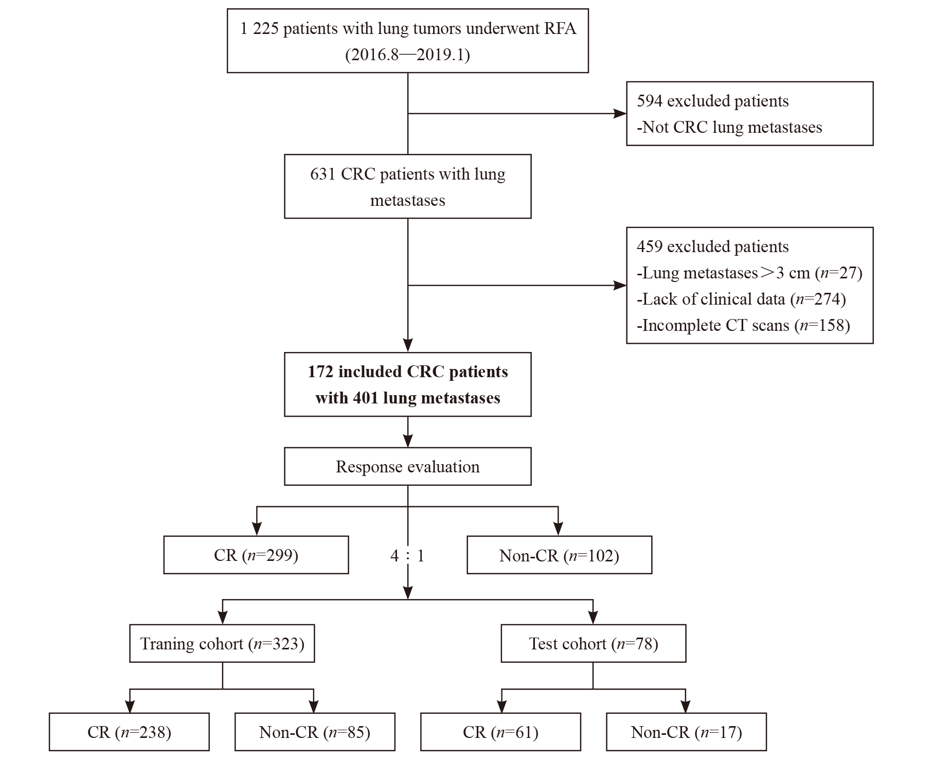
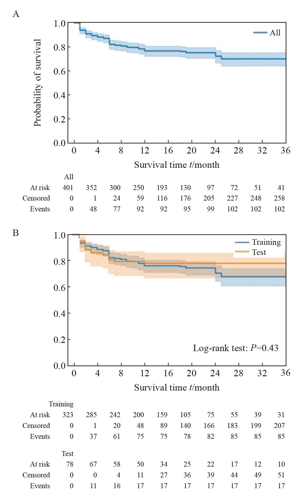
Fig. 3
Kaplan-Meier curve of LTPFS after RFA in CRC patients with lung metastases A: Kaplan-Meier curve of tumor recurrence after RFA in all patients; B: Kaplan-Meier curves of tumor recurrence after RFA in patients in the training and test datasets."
Tab. 2
Univariate and multivariate COX regression analysis of clinical and radiogical features"
| Characteristic | Univariate analysis | Multivariate analysis | |||
|---|---|---|---|---|---|
| HR (95% CI) | P value | HR (95% CI) | P value | ||
| Clinical features | |||||
| Gender | |||||
| Male | 1.055 (0.687-1.620) | 0.806 | |||
| Female | 1 | ||||
| Age | 1.003 (0.983-1.023) | 0.748 | |||
| Initial tumor location | |||||
| Rectum | 1 | ||||
| Sigmoid-left colon | 0.663 (0.318-1.383) | 0.273 | |||
| Transverse-right colon | 0.950 (0.532-1.695) | 0.862 | |||
| Caecum* | - | - | |||
| Lymphadenopathy at diagnosis | |||||
| Yes | 1.034 (0.659-1.621) | 0.884 | |||
| No | 1 | ||||
| Systemic therapy | |||||
| Yes | 0.817 (0.524-1.275) | 0.374 | |||
| No | 1 | ||||
| Tumor biomarkers | |||||
| CEA/(ng·mL-1) | 1.004 (1.001-1.007) | 0.002 | 1.052 (1.021-1.084) | 0.001 | |
| CA 19-9/(U·mL-1) | 1.003 (1.000-1.007) | 0.062 | 1.047 (1.008-1.088) | 0.019 | |
| Pre-RFA features of the lung metastases | |||||
| Location | |||||
| RUL | 1.035 (0.506-2.112) | 0.925 | 1.327 (0.660-2.668) | 0.427 | |
| RML | 1.612 (0.723-3.594) | 0.243 | 1.645 (0.760-3.564) | 0.207 | |
| RLL | 2.588 (1.358-4.929) | 0.004 | 3.055 (1.629-5.732) | 0.001 | |
| LUL | 1 | 1 | |||
| LLL | 1.732 (0.892-3.363) | 0.104 | 2.236 (1.192-4.193) | 0.012 | |
| Distance 1/cm | |||||
| ≥1 | 1 | ||||
| <1 | 1.326 (0.789-2.230) | 0.287 | |||
| Distance 2/cm | |||||
| ≥1 | 1 | ||||
| <1 | 1.206 (0.774-1.879) | 0.409 | |||
| Immediate post-RFA features | |||||
| Pneumothorax | |||||
| Yes | 1.207 (0.758-1.923) | 0.428 | |||
| No | 1 | ||||
| Hemorrhage | |||||
| Yes | 0.812 (0.492-1.339) | 0.414 | |||
| No | 1 | ||||
| Electrode | |||||
| Expandable | 0.480 (0.240-0.958) | 0.037 | 2.171 (1.176-4.005) | 0.013 | |
| Straight | 1 | 1 | |||
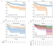
Fig. 4
Kaplan-Meier curves according to independent risk factors A: The risk stratification curves of CEA (optimal cutoff value was 6.28 ng/mL); B: The risk stratification curves of CA19-9 (optimal cutoff value was 74.72 U/mL); C: The risk stratification curves of ablation probe type; D: The risk stratification curves of position of lung metastases."

Tab. 3
Radiomics features screened by MRMRA and LASSO regression"
| Pre-RFA radiomics feature | Post-RFA radiomics feature |
|---|---|
| Shape_Elongation | Shape_Sphericity |
| Shape_MeshVolume | GLCM_Idn* |
| FirstOrder_Energy* | GLCM_Idmn |
| FirstOrder_Entropy* | GLDM_SmallDependenceLowGrayLevelEmphasis* |
| FirstOrder_MeanAbsoluteDeviation* | GLSZM_HighGrayLevelZoneEmphasis |
| FirstOrder_Skewness | NGTDM_Strength |
| FirstOrder_RootMeanSquared | NGTDM_Coarseness* |
| FirstOrder_RobustMeanAbsoluteDeviation | |
| GLCM_ClusterShade* | |
| GLCM_ClusterProminence | |
| GLCM_Idmn | |
| GLCM_Imc1* | |
| GLCM_InverseVariance* | |
| GLDM_DependenceEntropy* | |
| GLDM_GrayLevelNonUniformity* | |
| GLSZM_LargeAreaEmphasis | |
| GLSZM_LargeAreaLowGrayLevelEmphasis* |
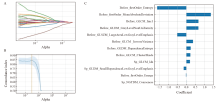
Fig. 5
Radiomics features screened by LASSO regression A: The coefficient of radiomic features changes with α. B: The C-index of the model changes with α, and the yellow line represents the α value corresponding to the highest value of the C-index in the cross-validation results. C: The correlation coefficient of radiomic features screened by LASSO regression. The prefix “before_”: The radiomic features of the preoperative CT images of RFA; The prefix “sp_”: The radiomics features of the CT images immediately after RFA."

Tab. 4
Performance comparison of each model"
| Variable | Fusion model | Radiomic model | Clinical model |
|---|---|---|---|
| Training dataset | |||
| C-index (95% CI) | 0.890 (0.854-0.927) | 0.867 (0.829-0.906) | 0.665 (0.594-0.737) |
| Brier score (1-year) | 0.092 | 0.106 | 0.161 |
| Brier score (2-year) | 0.160 | 0.189 | 0.180 |
| Brier score (3-year) | 0.161 | 0.198 | 0.177 |
| AUC (1-year) | 0.917 | 0.899 | 0.670 |
| AUC (2-year) | 0.869 | 0.850 | 0.668 |
| AUC (3-year) | 0.857 | 0.839 | 0.659 |
| Test dataset | |||
| C-index (95% CI) | 0.843 (0.768-0.916) | 0.811 (0.722-0.897) | 0.688 (0.565-0.811) |
| Brier score (1-year) | 0.126 | 0.133 | 0.172 |
| Brier score (2-year) | 0.204 | 0.241 | 0.191 |
| Brier score (3-year) | 0.174 | 0.188 | 0.187 |
| AUC (1-year) | 0.857 | 0.810 | 0.711 |
| AUC (2-year) | 0.709 | 0.618 | 0.687 |
| AUC (3-year) | 0.757 | 0.674 | 0.655 |
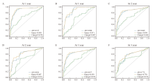
Fig. 6
ROC curves and AUC of each model for predicting LTPFS The ROC curves of each model predicted 1-year, 2-year, and 3-year LTPFS rates in the training set (A, C, E) and the test set (B, D, F), respectively. All: Fusion model; Omics: Radiomics model; Clinical: Clinical model."
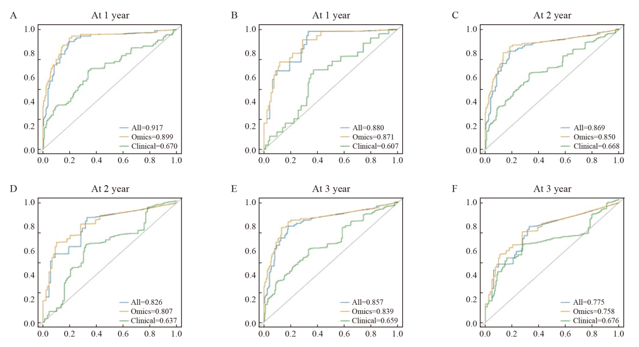
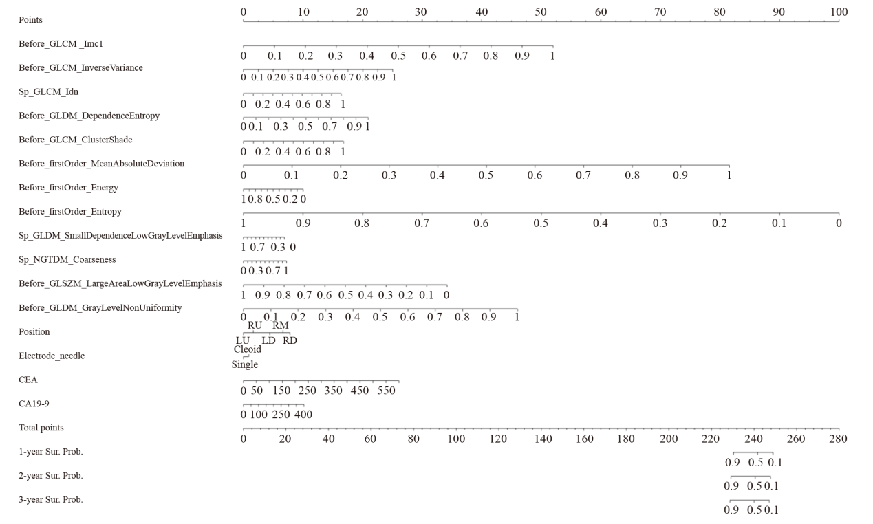
Fig. 7
Nomogram based on clinical variables and radiomics features"
| [1] | SUNG H, FERLAY J, SIEGEL R L, et al. Global cancer statistics 2020: GLOBOCAN estimates of incidence and mortality worldwide for 36 cancers in 185 countries[J]. CA Cancer J Clin, 2021, 71(3): 209-249. |
| [2] | NORDHOLM-CARSTENSEN A, KRARUP P M, JORGENSEN L N, et al. Occurrence and survival of synchronous pulmonary metastases in colorectal cancer: a nationwide cohort study[J]. Eur J Cancer, 2014, 50(2): 447-456. |
| [3] | SIEGEL R L, MILLER K D, JEMAL A. Cancer statistics, 2019[J]. CA, 2019, 69(1): 7-34. |
| [4] |
CHO J H, KIM S, NAMGUNG M, et al. The prognostic importance of the number of metastases in pulmonary metastasectomy of colorectal cancer[J]. World J Surg Oncol, 2015, 13: 222.
doi: 10.1186/s12957-015-0621-7 pmid: 26205014 |
| [5] |
IBRAHIM T, TSELIKAS L, YAZBECK C, et al. Systemic versus local therapies for colorectal cancer pulmonary metastasis: what to choose and when?[J]. J Gastrointest Cancer, 2016, 47(3): 223-231.
doi: 10.1007/s12029-016-9818-4 pmid: 27080402 |
| [6] |
QI H, FAN W J. Value of ablation therapy in the treatment of lung metastases[J]. Thorac Cancer, 2018, 9(2): 199-207.
doi: 10.1111/1759-7714.12567 pmid: 29193688 |
| [7] |
YU W S, BAE M K, CHOI J K, et al. Pulmonary metastasectomy in colorectal cancer: a population-based retrospective cohort study using the Korean national health insurance database[J]. Cancer Res Treat, 2021, 53(4): 1104-1112.
doi: 10.4143/crt.2020.1213 pmid: 33494126 |
| [8] |
NAJAFI A, BAERE T D, PURENNE E, et al. Risk factors for local tumor progression after RFA of pulmonary metastases: a matched case-control study[J]. Eur Radiol, 2021, 31(7): 5361-5369.
doi: 10.1007/s00330-020-07675-y pmid: 33474569 |
| [9] | CERVANTES A, ADAM R, ROSELLÓ S, et al. Metastatic colorectal cancer: ESMO clinical practice guideline for diagnosis, treatment and follow-up[J]. Ann Oncol, 2023, 34(1): 10-32. |
| [10] |
YANG Q X, QI H, ZHANG R, et al. Risk factors for local progression after percutaneous radiofrequency ablation of lung tumors: evaluation based on a review of 147 tumors[J]. J Vasc Interv Radiol, 2017, 28(4): 481-489.
doi: S1051-0443(16)30842-9 pmid: 28111196 |
| [11] | DOO F X, VOSSHENRICH J, COOK T S, et al. Environmental sustainability and AI in radiology: a double-edged sword[J]. Radiology, 2024, 310(2): e232030. |
| [12] |
BRANCATO V, ESPOSITO G, COPPOLA L, et al. Standardizing digital biobanks: Integrating imaging, genomic, and clinical data for precision medicine[J]. J Transl Med, 2024, 22(1): 136.
doi: 10.1186/s12967-024-04891-8 pmid: 38317237 |
| [13] | TERRANOVA N, VENKATAKRISHNAN K. Machine learning in modeling disease trajectory and treatment outcomes: an emerging enabler for model-informed precision medicine[J]. Clin Pharmacol Ther, 2024, 115(4): 720-726. |
| [14] |
BEIG N, KHORRAMI M, ALILOU M, et al. Perinodular and intranodular radiomic features on lung CT images distinguish adenocarcinomas from granulomas[J]. Radiology, 2019, 290(3): 783-792.
doi: 10.1148/radiol.2018180910 pmid: 30561278 |
| [15] |
ABTIN F G, ERADAT J, GUTIERREZ A J, et al. Radiofrequency ablation of lung tumors: imaging features of the postablation zone[J]. Radiographics, 2012, 32(4): 947-969.
doi: 10.1148/rg.324105181 pmid: 22786987 |
| [16] | 刘宝东, 支修益. 影像引导下热消融治疗肺部肿瘤的局部疗效评价[J]. 中国医学前沿杂志(电子版), 2015, 7(2): 11-14. |
| [17] | LIU B D, ZHI X Y. Evaluation of local curative effect of image-guided thermal ablation for lung tumors[J]. Chin J Front Med Sci Electron Version, 2015, 7(2): 11-14. |
| [18] | STARMANS M P A, VAN DER VOORT S R, PHIL T, et al. Reproducible radiomics through automated machine learning validated on twelve clinical applications[EB/OL]. (2022-07-29) [2024-05-07]. http://arxiv.org/abs/2108.08618 |
| [19] |
RIGATTI S J. Random forest[J]. J Insur Med, 2017, 47(1): 31-39.
doi: 10.17849/insm-47-01-31-39.1 pmid: 28836909 |
| [20] | QIAN L Q, ZHOU Y J, ZENG W Q, et al. A random forest algorithm predicting model combining intraoperative frozen section analysis and clinical features guides surgical strategy for peripheral solitary pulmonary nodules[J]. Transl Lung Cancer Res, 2022, 11(6): 1132-1144. |
| [21] | KHUSHI M, SHAUKAT K, ALAM T M, et al. A comparative performance analysis of data resampling methods on imbalance medical data[J]. IEEE Access, 2021, 9: 109960-109975. |
| [22] | HALL D L, LLINAS J. An introduction to multisensor data fusion[J]. Proc IEEE, 1997, 85(1): 6-23. |
| [23] | RASHINKAR P, KRUSHNASAMY V S. An overview of data fusion techniques[C]. Bengaluru: ICIMIA, 2017: 694-697. |
| [24] |
MARKICH R, PALUSSIÈRE J, CATENA V, et al. Radiomics complements clinical, radiological, and technical features to assess local control of colorectal cancer lung metastases treated with radiofrequency ablation[J]. Eur Radiol, 2021, 31(11): 8302-8314.
doi: 10.1007/s00330-021-07998-4 pmid: 33954806 |
| [25] |
BAÈRE T D, AUPÉRIN A, DESCHAMPS F, et al. Radiofrequency ablation is a valid treatment option for lung metastases: experience in 566 patients with 1037 metastases[J]. Ann Oncol, 2015, 26(5): 987-991.
doi: S0923-7534(19)31500-5 pmid: 25688058 |
| [26] |
GONZALEZ M, PONCET A, COMBESCURE C, et al. Risk factors for survival after lung metastasectomy in colorectal cancer patients: a systematic review and meta-analysis[J]. Ann Surg Oncol, 2013, 20(2): 572-579.
doi: 10.1245/s10434-012-2726-3 pmid: 23104709 |
| [27] |
PALUSSI RE J, MARCET B, DESCAT E, et al. Lung tumors treated with percutaneous radiofrequency ablation: computed tomography imaging follow-up[J]. Cardiovasc Intervent Radiol, 2011, 34(5): 989-997.
doi: 10.1007/s00270-010-0048-z pmid: 21127867 |
| [28] | MAPELLI P, BEZZI C, MUFFATTI F, et al. Preoperative assessment of lymph nodal metastases with[68Ga]Ga-DOTATOC PET radiomics for improved surgical planning in well-differentiated pancreatic neuroendocrine tumours[J]. Eur J Nucl Med Mol Imaging, 2024, 51(9): 2774-2783. |
| [29] |
ZHU F D, YANG C, XIA Y, et al. CT-based radiomics models may predict the early efficacy of microwave ablation in malignant lung tumors[J]. Cancer Imaging, 2023, 23(1): 60.
doi: 10.1186/s40644-023-00571-w pmid: 37308918 |
| [30] |
HUANG H Z, CHEN H, ZHENG D Z, et al. Habitat-based radiomics analysis for evaluating immediate response in colorectal cancer lung metastases treated by radiofrequency ablation[J]. Cancer Imaging, 2024, 24(1): 44.
doi: 10.1186/s40644-024-00692-w pmid: 38532520 |
| [31] | BENSON A B, VENOOK A P, AL-HAWARY M M, et al. Colon cancer, version 2.2021, NCCN clinical practice guidelines in oncology[J]. J Natl Compr Canc Netw, 2021, 19(3): 329-359. |
| [32] |
DUFFY M J, LAMERZ R, HAGLUND C, et al. Tumor markers in colorectal cancer, gastric cancer and gastrointestinal stromal cancers: European group on tumor markers 2014 guidelines update[J]. Int J Cancer, 2014, 134(11): 2513-2522.
doi: 10.1002/ijc.28384 pmid: 23852704 |
| [33] | LIU B, LI C H, SUN X R, et al. Assessment and prognostic value of immediate changes in post-ablation intratumor density heterogeneity of pulmonary tumors via radiomics-based computed tomography features[J]. Front Oncol, 2021, 11: 615174. |
| Viewed | ||||||
|
Full text |
|
|||||
|
Abstract |
|
|||||
沪ICP备12009617
Powered by Beijing Magtech Co. Ltd