Welcome to China Oncology,
China Oncology ›› 2023, Vol. 33 ›› Issue (6): 597-604.doi: 10.19401/j.cnki.1007-3639.2023.06.007
• Article • Previous Articles Next Articles
DU Shuxiang1( ), ZHAO Yingzhi1, ZHANG Xuetao1, WANG Tao1, LUO Jingyu2, ZHANG Wei1, WU Gang1()
Received:2022-08-10
Revised:2023-03-14
Online:2023-06-30
Published:2023-07-26
Share article
CLC Number:
DU Shuxiang, ZHAO Yingzhi, ZHANG Xuetao, WANG Tao, LUO Jingyu, ZHANG Wei, WU Gang. Analysis of group 12a lymph node metastasis at different sites of gastric adenocarcinoma and its risk factors[J]. China Oncology, 2023, 33(6): 597-604.

Fig. 1
Lymph node dissection steps in group No.12a GDA: The gastroduodenal artery; RGA: The right gastric artery; CHA: The common hepatic artery; PHA: The proper hepatic artery; PV: The portal vein. A: Severed the right gastroomentum artery, free the gastroduodenal artery; B: Bared the common hepatic artery, the right gastric artery and the proper hepatic artery; C: Exposed the portal vein, separate along the front of the portal vein, and clean the lymph nodes between the portal vein and the liver intrinsic artery; D: Lifted down the intrinsic artery of the liver to sweep away the surrounding adipose tissue; E: Opened the hepatoduodenal ligament and clean the 12a group lymph nodes; F: Lymph nodes were picked in groups within 30minutes after the specimen is left."
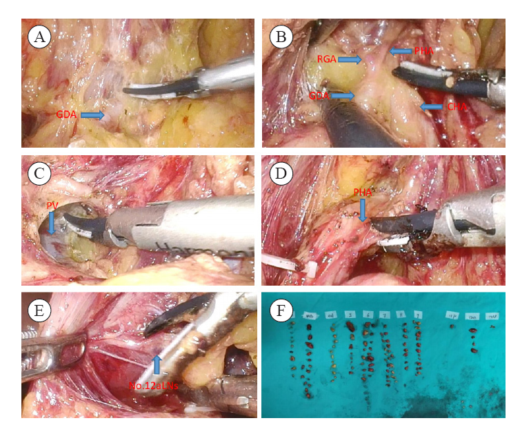

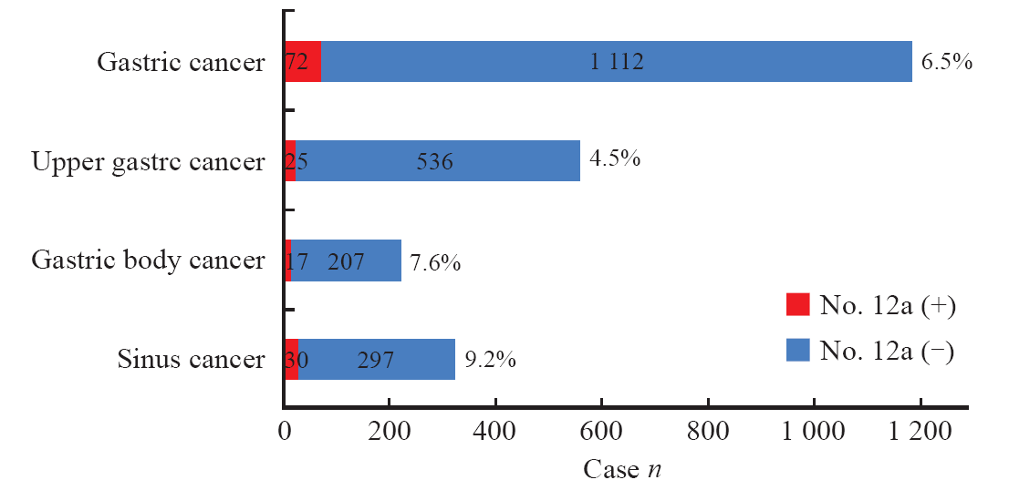
Fig. 2
Lymph node metastasis rate of the group 12a lymph node in each location"
Tab. 1
The subgroup analysis and univariate analysis of No.12a lymph node metastasis in gastric cancer in each location [n (%)]"
| Clinicopathological data | Upper cancer (n=561) | Body cancer (n=224) | Antral cancer (n=327) | |||||||||||
|---|---|---|---|---|---|---|---|---|---|---|---|---|---|---|
| No.12a (+) (n=25) | No.12a (-) (n=536) | χ2 | P value | No.12a (+) (n=17) | No.12a (-) (n=207) | χ2 | P value | No.12a (+) (n=30) | No.12a (-) (n=297) | χ2 | P value | |||
| Gender | 0.841 | 0.359 | 0.829 | 0.362 | 0.984 | 0.321 | ||||||||
| Male | 16 (5.2) | 293 (94.8) | 7 (9.3) | 109 (90.7) | 19 (10.6) | 160 (89.4) | ||||||||
| Female | 9 (3.6) | 243 (96.4) | 10 (9.2) | 98 (90.8) | 11 (7.4) | 137 (92.6) | ||||||||
| Age/year | 0.066 | 0.789 | 0.200 | 0.654 | 0.066 | 0.767 | ||||||||
| <65 | 14 (4.3) | 314 (95.7) | 10 (7.0) | 133 (93.0) | 13 (8.7) | 136 (91.3) | ||||||||
| ≥65 | 11 (4.7) | 222 (95.3) | 7 (8.6) | 74 (91.4) | 17 (9.6) | 161 (90.4) | ||||||||
| BMI/(kg·m-2) | 5.562 | 0.018 | 3.100 | 0.078 | 14.064 | <0.001 | ||||||||
| <24 | 14 (3.3) | 411 (96.7) | 16 (9.3) | 156 (90.7) | 13 (5.8) | 224 (94.2) | ||||||||
| ≥24 | 11 (8.1) | 125 (91.9) | 1 (1.9) | 51 (98.2) | 17 (18.9) | 73 (81.1) | ||||||||
| Tumor size D/cm | 155.058 | <0.001 | 90.044 | <0.001 | 137.080 | <0.001 | ||||||||
| <6 | 0 (0.0) | 480 (100.0) | 0 (0.0) | 186 (100.0) | 6 (2.1) | 280 (97.9) | ||||||||
| ≥6 | 25 (30.9) | 56 (69.1) | 17 (44.7) | 21 (55.3) | 24 (58.5) | 17 (41.5) | ||||||||
| Borrmann‘s classification | 1.351 | 0.509 | 0.112 | 0.946 | 4.999 | 0.082 | ||||||||
| Uplift | 2 (4.9) | 39 (95.1) | 1 (8.3) | 11 (91.7) | 1 (5.6) | 17 (94.4) | ||||||||
| Ulcer | 20 (4.1) | 466 (95.9) | 15 (9.3) | 150 (90.7) | 25 (8.4) | 273 (91.6) | ||||||||
| Infiltrating | 3 (8.8) | 31(91.2) | 1 (6.7) | 14 (93.3) | 4 (9.3) | 7 (90.7) | ||||||||
| Differentiation | 0.325 | 0.569 | 0.888 | 0.346 | 3.545 | 0.060 | ||||||||
| Poorly differentiated | 4 (3.5) | 111(96.5) | 5 (10.9) | 41 (89.1) | 10 (15.2) | 56 (84.8) | ||||||||
| Medium to high differentiation | 21 (4.7) | 425 (95.3) | 12(6.7) | 166(93.3) | 20 (7.7) | 241 (92.3) | ||||||||
| pT staging | 0.454 | 0.011 | 8.362 | 0.004 | 13.167 | <0.001 | ||||||||
| T1-T2 | 0 (0.0) | 111 (100.0) | 0 (0.0) | 70 (100.0) | 1 (9.3) | 107 (90.7) | ||||||||
| T3-T4 | 25(5.6) | 425 (94.4) | 17(11.0) | 137 (99.0) | 29 (13.2) | 190 (86.8) | ||||||||
| pN staging | 39.075 | <0.001 | 4.303 | 0.038 | 88.328 | <0.001 | ||||||||
| N0-N1 | 0 (0.0) | 336 (100.0) | 0 (0.0) | 123 (100.0) | 0 (0.0) | 238 (100.0) | ||||||||
| N2-N3 | 25 (11.1) | 200 (88.9) | 3 (3.4) | 84 (96.6) | 30 (33.7) | 59 (66.3) | ||||||||
| TNM staging | 20.251 | <0.001 | 9.330 | 0.002 | 7.882 | 0.005 | ||||||||
| Ⅰ-Ⅱ | 1 (0.4) | 268 (99.6) | 2 (1.9) | 104 (98.1) | 0 (0.0) | 63 (100.0) | ||||||||
| Ⅲ-Ⅳ | 24 (8.2) | 268 (91.8) | 15 (12.7) | 103 (87.3) | 30 (11.4) | 234 (88.6) | ||||||||
| Neurovascular invasion | 0.067 | 0.769 | 10.695 | 0.001 | 2.384 | 0.123 | ||||||||
| Yes | 14 (4.7) | 286 (95.3) | 8 (20.0) | 32 (80.0) | 14 (12.6) | 97 (87.4) | ||||||||
| No | 11 (4.2) | 250 (95.8) | 9 (4.9) | 175 (95.1) | 16 (7.4) | 200 (92.6) | ||||||||
Tab. 2
Variable assignment table"
| Variable | Assignment table |
|---|---|
| Dependent variable | No.12a lymph node metastasis was 1; Lymph node metastasis in No.12a was 0 |
| Independent variable | |
| BMI | ≥24 kg/m2 was“1”, <24 kg/m² was “0” |
| Tumor size | ≥6 cm was “1”, <6 cm was “0” |
| pT staging | T1 was “1”, T2 was “2”, T3 was “3”, T4 was “4” |
| TNM staging | Ⅰ was “1”, Ⅱ was “2”, Ⅲ was “3” |
| Vascular nerve invasion | “Yes” was “1”,“No”was“0” |
Tab. 3
Multivariable logistic regression analysis of risk factors for No.12a lymph node metastasis in upper gastric cancer"
| Variable | β | SE | Wald χ2 | P value | OR | 95% CI |
|---|---|---|---|---|---|---|
| BMI | -0.050 | 0.030 | 2.844 | 0.092 | 0.951 | 0.898-1.008 |
| Tumor size | 1.139 | 0.038 | 877.688 | <0.001 | 3.122 | 2.896-3.366 |
| pT staging | 0.878 | 0.176 | 24.982 | <0.001 | 2.406 | 1.705-3.396 |
| TNM staging | 2.068 | 0.260 | 63.415 | <0.001 | 7.910 | 4.755-13.160 |
Tab. 4
Multivariable logistic regression analysis of risk factors for No.12a lymph node metastasis in gastric antral cancer"
| Variable | β | SE | Wald χ2 | P value | OR | 95% CI |
|---|---|---|---|---|---|---|
| Tumor size | 1.014 | 0.051 | 394.733 | <0.001 | 2.757 | 2.495-3.047 |
| pT staging | 1.193 | 0.174 | 47.112 | <0.001 | 3.298 | 2.346-4.638 |
| TNM staging | 0.164 | 0.189 | 0.747 | 0.388 | 1.178 | 0.813-1.707 |
| Vascular nerve invasion | 0.609 | 0.151 | 16.275 | <0.001 | 1.839 | 1.368-2.473 |
Tab. 5
Multivariable logistic regression analysis of risk factors for No.12a lymph node metastasis in gastric body cancer group"
| Variable | β | SE | Wald χ2 | P value | OR | 95% CI |
|---|---|---|---|---|---|---|
| BMI | 0.093 | 0.036 | 6.691 | 0.01 | 1.098 | 1.023-1.178 |
| Tumor size | 1.053 | 0.243 | 18.708 | <0.001 | 2.865 | 1.778-4.616 |
| pT staging | 2.602 | 0.244 | 113.565 | <0.001 | 13.497 | 8.363-21.783 |
| TNM staging | 1.337 | 0.103 | 169.942 | <0.001 | 3.806 | 3.113-4.653 |
| [1] |
SUNG H, FERLAY J, SIEGEL R L, et al. Global cancer statistics 2020: GLOBOCAN estimates of incidence and mortality worldwide for 36 cancers in 185 countries[J]. CA Cancer J Clin, 2021, 71(3): 209-249.
doi: 10.3322/caac.v71.3 |
| [2] | 李国新. 胃癌外科2021年研究进展及学科展望[J]. 中华胃肠外科杂志, 2022, 25(1): 15-21. |
| LI G X. Research progress and prospect of gastric cancer surgery in 2021[J]. Chin J Gastrointest Surg, 2022, 25(1): 15-21. | |
| [3] |
DONG Y P, CAI F L, WU Z Z, et al. Risk of station 12a lymph node metastasis in patients with lower-third gastric cancer[J]. World J Gastrointest Surg, 2021, 13(11): 1390-1404.
doi: 10.4240/wjgs.v13.i11.1390 |
| [4] |
SIERRA A, REGUEIRA F M, HERNÁNDEZ-LIZOÁIN J L, et al. Role of the extended lymphadenectomy in gastric cancer surgery: experience in a single institution[J]. Ann Surg Oncol, 2003, 10(3): 219-226.
pmid: 12679305 |
| [5] |
JAPANESE GASTRIC CANCER ASSOCIATION. Japanese gastric cancer treatment guidelines 2018 (5th edition)[J]. Gastric Cancer, 2021, 24(1): 1-21.
doi: 10.1007/s10120-020-01042-y |
| [6] |
JAPANESE GASTRIC CANCER ASSOCIATION. Japanese classification of gastric carcinoma-2nd English edition[J]. Gastric Cancer, 1998, 1(1): 10-24.
doi: 10.1007/PL00011681 |
| [7] |
EDGE S B, COMPTON C C. The American Joint Committee on Cancer: the 7th edition of the AJCC cancer staging manual and the future of TNM[J]. Ann Surg Oncol, 2010, 17(6): 1471-1474.
doi: 10.1245/s10434-010-0985-4 pmid: 20180029 |
| [8] |
LU J, ZHENG C H, CAO L L, et al. The effectiveness of the 8th American Joint Committee on Cancer TNM classification in the prognosis evaluation of gastric cancer patients: a comparative study between the 7th and 8th editions[J]. Eur J Surg Oncol, 2017, 43(12): 2349-2356.
doi: S0748-7983(17)30659-5 pmid: 28943179 |
| [9] |
HARTGRINK H H, VAN DE VELDE C J, PUTTER H, et al. Extended lymph node dissection for gastric cancer: who may benefit? Final results of the randomized Dutch gastric cancer group trial[J]. J Clin Oncol, 2004, 22(11): 2069-2077.
doi: 10.1200/JCO.2004.08.026 pmid: 15082726 |
| [10] |
CUSCHIERI A, WEEDEN S, FIELDING J, et al. Patient survival after D1 and D2 resections for gastric cancer: long-term results of the MRC randomized surgical trial. Surgical Co-operative Group[J]. Br J Cancer, 1999, 79(9/10): 1522-1530.
doi: 10.1038/sj.bjc.6690243 |
| [11] |
KONG S H, YOO M W, KIM J W, et al. Validation of limited lymphadenectomy for lower-third gastric cancer based on depth of tumour invasion[J]. Br J Surg, 2011, 98(1): 65-72.
doi: 10.1002/bjs.7266 pmid: 20954197 |
| [12] | WEI Z W, XIA G K, WU Y, et al. Evaluation of skeletonization of the hepatoduodenal ligament for the lower third gastric cancer by propensity score analysis[J]. Hepatogastroenterology, 2013, 60(127): 1789-1796. |
| [13] |
CAI S R, CHEN J H, CHEN C Q, et al. Survival of proper hepatic artery lymph node metastasis in patients with gastric cancer: implications for D2 lymphadenectomy[J]. PLoS One, 2015, 10(3): e0118953.
doi: 10.1371/journal.pone.0118953 |
| [14] |
KUMAGAI K, HIKI N, NUNOBE S, et al. Metastasis to the lymph nodes along the proper hepatic artery from adenocarcinoma of the stomach[J]. Langenbecks Arch Surg, 2016, 401(5): 677-685.
doi: 10.1007/s00423-016-1429-9 |
| [15] |
IN H, SOLSKY I, PALIS B, et al. Validation of the 8th edition of the AJCC TNM staging system for gastric cancer using the national cancer database[J]. Ann Surg Oncol, 2017, 24(12): 3683-3691.
doi: 10.1245/s10434-017-6078-x pmid: 28895113 |
| [16] |
CHEN Z H, LIU Y, DOU L Z, et al. The efficacy of the application of the curative criteria of the 5th edition Japanese gastric cancer treatment guidelines for early adenocarcinoma of the esophagogastric junction treated by endoscopic submucosal dissection[J]. Saudi J Gastroenterol, 2021, 27(2): 97-104.
doi: 10.4103/sjg.SJG_403_20 |
| [17] |
AJANI J A, D’AMICO T A, BENTREM D J, et al. Gastric cancer, version 2.2022, NCCN clinical practice guidelines in oncology[J]. J Natl Compr Canc Netw, 2022, 20(2): 167-192.
doi: 10.6004/jnccn.2022.0008 |
| [18] |
SMITH D D, SCHWARZ R R, SCHWARZ R E. Impact of total lymph node count on staging and survival after gastrectomy for gastric cancer: data from a large US-population database[J]. J Clin Oncol, 2005, 23(28): 7114-7124.
doi: 10.1200/JCO.2005.14.621 pmid: 16192595 |
| [19] |
SCHWARZ R E, SMITH D D. Clinical impact of lymphadenectomy extent in resectable gastric cancer of advanced stage[J]. Ann Surg Oncol, 2007, 14(2): 317-328.
doi: 10.1245/s10434-006-9218-2 pmid: 17094022 |
| [20] | SEEVARATNAM R, BOCICARIU A, CARDOSO R, et al. How many lymph nodes should be assessed in patients with gastric cancer? A systematic review[J]. Gastric Cancer, 2012, 15(Suppl 1): S70-S88. |
| [21] | 张晋杰, 王杰, 胡文庆. 进展期胃上部癌淋巴结清扫的现状与研究进展[J]. 中华胃肠外科杂志, 2018, 21(2): 236-240. |
| ZHANG J J, WANG J, HU W Q. Current status and research progress of lymph node dissection in advanced upper gastric cancer[J]. Chin J Gastrointest Surg, 2018, 21(2): 236-240. | |
| [22] | 郑民华, 臧潞, 马君俊, 等. SiewertⅡ型食管胃结合部腺癌腔镜手术治疗中国专家共识(2019版)[J]. 中国实用外科杂志, 2019, 39(11): 1129-1135. |
| ZHENG M H, ZANG L, MA J J, et al. Endoscopic surgery for SiewertⅡ esophagogastric junction adenocarcinoma in China (2019 edition)[J]. Chin J Pract Surg, 2019, 39(11): 1129-1135. | |
| [23] |
SHU P, SUN X F, LIU F L, et al. Pattern of No. 12a lymph node metastasis in gastric cancer[J]. Chin J Cancer Res, 2021, 33(1): 61-68.
doi: 10.21147/j.issn.1000-9604.2021.01.07 |
| [24] |
SATO Y, KATAI H, ITO M, et al. Can proximal gastrectomy be justified for advanced adenocarcinoma of the esophagogastric junction?[J]. J Gastric Cancer, 2018, 18(4): 339-347.
doi: 10.5230/jgc.2018.18.e33 pmid: 30607297 |
| [25] |
WU G, ZHANG D Y, DUAN Y H, et al. Correlations of hemoglobin level and perioperative blood transfusion with the prognosis of gastric cancer: a retrospective study[J]. Med Sci Monit, 2017, 23: 2470-2478.
doi: 10.12659/MSM.900907 |
| [26] |
MITA K, ITO H, KATSUBE T, et al. Prognostic factors affecting survival after multivisceral resection in patients with clinical T4b gastric cancer[J]. J Gastrointest Surg, 2017, 21(12): 1993-1999.
doi: 10.1007/s11605-017-3559-y |
| [27] |
YAMASHITA H, KATAI H, MORITA S, et al. Optimal extent of lymph node dissection for Siewert type Ⅱ esophagogastric junction carcinoma[J]. Ann Surg, 2011, 254(2): 274-280.
doi: 10.1097/SLA.0b013e3182263911 |
| Viewed | ||||||
|
Full text |
|
|||||
|
Abstract |
|
|||||
沪ICP备12009617
Powered by Beijing Magtech Co. Ltd