Welcome to China Oncology,
China Oncology ›› 2025, Vol. 35 ›› Issue (1): 49-57.doi: 10.19401/j.cnki.1007-3639.2025.01.006
• Specialist' Commentary • Previous Articles Next Articles
WANG Renfei( ), LU Gaixia
Received:2024-12-13
Revised:2025-01-21
Online:2025-01-30
Published:2025-02-17
Contact:
WANG Renfei
Share article
CLC Number:
WANG Renfei, LU Gaixia. The unique value and controversy of nuclear medicine molecular imaging in the evaluation of radioiodine-refractory differentiated thyroid cancer[J]. China Oncology, 2025, 35(1): 49-57.
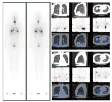
Fig. 1
131I-WBS and SPECT/CT"
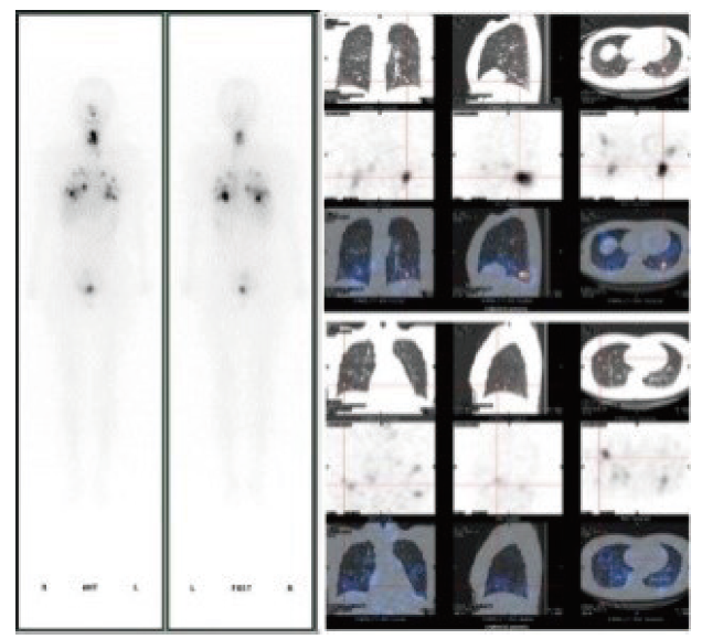

Fig. 2
Negative 131I-WBS and positive 18F-FDG PET/CT"
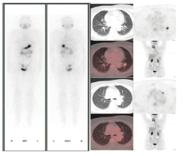
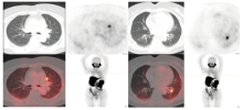
Fig. 3
18F-PSMA PET/CT"
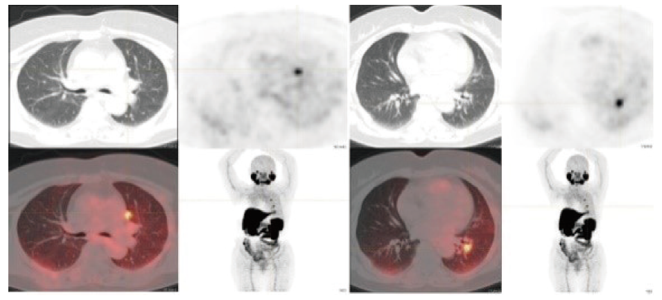
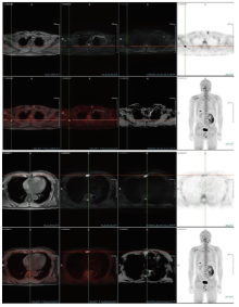
Fig. 4
18F-FAPI PET/MRI"

| [1] | HAUGEN B R, ALEXANDER E K, BIBLE K C, et al. 2015 American thyroid association management guidelines for adult patients with thyroid nodules and differentiated thyroid cancer: the American Thyroid Association guidelines task force on thyroid nodules and differentiated thyroid cancer[J]. Thyroid, 2016, 26(1): 1-133. |
| [2] | 中国临床肿瘤学会核医学专家委员会, 中国临床肿瘤学会甲状腺癌专家委员会, 中华医学会核医学分会, 等. 放射性碘难治性分化型甲状腺癌诊治管理指南(2024版)[J]. 中华核医学与分子影像杂志, 2024, 44(6): 359-372. |
| Nuclear Medicine Expert Committee of Chinese Society of Clinical Oncology, Thyroid Cancer Expert Committee of Chinese Society of Clinical Oncology, Thyroid Cancer Expert Committee of Chinese Society of Clinical Oncology, Chinese Society of Nuclear Medicine, et al. Management guidelines for radioactive iodine-refractory differentiated thyroid cancer (2024 edition)[J]. Chin J Nucl Med Mol Imag, 2024, 44(6): 359-372. | |
| [3] |
JIN Y C, VAN NOSTRAND D, CHENG L X, et al. Radioiodine refractory differentiated thyroid cancer[J]. Crit Rev Oncol Hematol, 2018, 125: 111-120.
doi: S1040-8428(17)30331-1 pmid: 29650270 |
| [4] | ZILIOLI V, PELI A, PANAROTTO M B, et al. Differentiated thyroid carcinoma: incremental diagnostic value of 131I SPECT/CT over planar whole body scan after radioiodine therapy[J]. Endocrine, 2017, 56(3): 551-559. |
| [5] | MORRIS L F, WAXMAN A D, BRAUNSTEIN G D. The nonimpact of thyroid stunning: remnant ablation rates in 131I-scanned and nonscanned individuals[J]. J Clin Endocrinol Metab, 2001, 86(8): 3507-3511. |
| [6] |
YAP B K, MURBY B. No adverse affect in clinical outcome using low preablation diagnostic (131)I activity in differentiated thyroid cancer: refuting thyroid-stunning effect[J]. J Clin Endocrinol Metab, 2014, 99(7): 2433-2440.
doi: 10.1210/jc.2014-1405 pmid: 24762114 |
| [7] | DE LA VIEJA A, RIESCO-EIZAGUIRRE G. Radio-iodide treatment: from molecular aspects to the clinical view[J]. Cancers (Basel), 2021, 13(5): 995. |
| [8] | SABRA M M, GREWAL R K, TALA H, et al. Clinical outcomes following empiric radioiodine therapy in patients with structurally identifiable metastatic follicular cell-derived thyroid carcinoma with negative diagnostic but positive post-therapy 131I whole-body scans[J]. Thyroid, 2012, 22(9): 877-883. |
| [9] |
SOUZA ROSÁRIO P W, BARROSO A L, REZENDE L L, et al. Post I-131 therapy scanning in patients with thyroid carcinoma metastases: an unnecessary cost or a relevant contribution?[J]. Clin Nucl Med, 2004, 29(12): 795-798.
pmid: 15545881 |
| [10] |
FATOURECHI V, HAY I D, MULLAN B P, et al. Are posttherapy radioiodine scans informative and do they influence subsequent therapy of patients with differentiated thyroid cancer?[J]. Thyroid, 2000, 10(7): 573-577.
doi: 10.1089/thy.2000.10.573 pmid: 10958309 |
| [11] | 王任飞, 高再荣, 欧阳伟, 等. 分化型甲状腺癌转移灶摄碘能力与131I清灶疗效关系的回顾性多中心研究[J]. 中华核医学与分子影像杂志, 2020, 40(6): 334-338. |
| WANG R F, GAO Z R, OUYANG W, et al. Correlation between 131I uptake and therapeutic efficacy in metastatic differentiated thyroid carcinoma: a retrospective multicenter study\n[J]. Chin J Nucl Med Mol Imag, 2020, 40(6): 334-338. | |
| [12] | SA R, CHENG L, JIN Y C, et al. Distinguishing patients with distant metastatic differentiated thyroid cancer who biochemically benefit from next radioiodine treatment[J]. Front Endocrinol (Lausanne), 2020, 11: 587315. |
| [13] |
GULEC S A, AHUJA S, AVRAM A M, et al. A joint statement from the American Thyroid Association, the European Association of Nuclear Medicine, the European Thyroid Association, the Society of Nuclear Medicine and Molecular Imaging on current diagnostic and theranostic approaches in the management of thyroid cancer[J]. Thyroid, 2021, 31(7): 1009-1019.
doi: 10.1089/thy.2020.0826 pmid: 33789450 |
| [14] | 中华医学会核医学分会. 131I治疗分化型甲状腺癌指南(2021版)[J]. 中华核医学与分子影像杂志, 2021, 41(4): 218-241. |
| Chinese Society of Nuclear Medicine. Guidelines for radioiodine therapy of differentiated thyroid cancer (2021 edition)[J]. Chin J Nucl Med Mol Imag, 2021, 41(4): 218-241. | |
| [15] | 中国临床肿瘤学会甲状腺癌专业委员会, 中国研究型医院学会分子诊断专业委员会甲状腺癌学组, 医促会甲状腺疾病专业委员会核医学组, 等. 分化型甲状腺癌术后131I治疗前评估专家共识[J]. 中国癌症杂志, 2019, 29(10): 832-840. |
| Chinese Clinical Oncology Society Thyroid Cancer Professional Committee, Thyroid Cancer Sub-group of the Molecular Diagnosis Professional Committee of the Chinese Research Hospital Association, Nuclear Medicine Group of the Thyroid Disease Professional Committee of the China Association for the Promotion of Medical and Health Care, et al. Expert consensus on pre-treatment assessment of 131I therapy after differentiated thyroid cancer surgery[J]. China Oncol, 2019, 29(10): 832-840. | |
| [16] | WAN Q C, BAI L, ZHAO G G, et al. Diagnostic performance of 18F-FDG-PET/CT in DTC patients with thyroglobulin elevation and negative iodine scintigraphy: a meta-analysis[J]. Eur J Endocrinol, 2019, 181(2): 93-102. |
| [17] | ALBANO D, TULCHINSKY M, DONDI F, et al. Thyroglobulin doubling time offers a better threshold than thyroglobulin level for selecting optimal candidates to undergo localizing [18F] FDG PET/CT in non-iodine avid differentiated thyroid carcinoma[J]. Eur J Nucl Med Mol Imaging, 2021, 48(2): 461-468. |
| [18] | GIOVANELLA L, TRIMBOLI P, VERBURG F A, et al. Thyroglobulin levels and thyroglobulin doubling time independently predict a positive 18F-FDG PET/CT scan in patients with biochemical recurrence of differentiated thyroid carcinoma[J]. Eur J Nucl Med Mol Imaging, 2013, 40(6): 874-880. |
| [19] | WANG H X, DAI H Y, LI Q R, et al. Investigating 18F-FDG PET/CT parameters as prognostic markers for differentiated thyroid cancer: a systematic review[J]. Front Oncol, 2021, 11: 648658. |
| [20] | ALBANO D, DONDI F, MAZZOLETTI A, et al. Prognostic role of 2-[18F] FDG PET/CT metabolic volume parameters in patients affected by differentiated thyroid carcinoma with high thyroglobulin level, negative 131I wbs and positive 2-[18F]-FDG PET/CT[J]. Diagnostics, 2021, 11(12): 2189. |
| [21] | ROBBINS R J, WAN Q, GREWAL R K, et al. Real-time prognosis for metastatic thyroid carcinoma based on 2-[18F] fluoro-2-deoxy-D-glucose-positron emission tomography scanning[J]. J Clin Endocrinol Metab, 2006, 91(2): 498-505. |
| [22] | AHMADDY F, BURGARD C, BEYER L, et al. 18F-FDG-PET/CT in patients with advanced, radioiodine refractory thyroid cancer treated with lenvatinib[J]. Cancers (Basel), 2021, 13(2): 317. |
| [23] | RENDL G, SCHWEIGHOFER-ZWINK G, SORKO S, et al. Assessment of treatment response to lenvatinib in thyroid cancer monitored by F-18 FDG PET/CT using PERCIST 1.0, modified PERCIST and EORTC criteria-which one is most suitable?[J]. Cancers (Basel), 2022, 14(8): 1868. |
| [24] | VALERIO L, GUIDOCCIO F, GIANI C, et al. [18F]-FDG-PET/CT correlates with the response of radiorefractory thyroid cancer to lenvatinib and patient survival[J]. J Clin Endocrinol Metab, 2021, 106(8): 2355-2366. |
| [25] | ZHAO D, JIN X N, LI F, et al. Integrin αvβ3 imaging of radioactive iodine-refractory thyroid cancer using 99mTc-3PRGD2[J]. J Nucl Med, 2012, 53(12): 1872-1877. |
| [26] | PARIHAR A S, MITTAL B R, KUMAR R, et al. 68Ga-DOTA-RGD2 positron emission tomography/computed tomography in radioiodine refractory thyroid cancer: prospective comparison of diagnostic accuracy with 18F-FDG positron emission tomography/computed tomography and evaluation toward potential theranostics[J]. Thyroid, 2020, 30(4): 557-567. |
| [27] | CIAPPUCCINI R, SAGUET-RYSANEK V, GIFFARD F, et al. PSMA expression in differentiated thyroid cancer: association with radioiodine, 18FDG uptake, and patient outcome[J]. J Clin Endocrinol Metab, 2021, 106(12): 3536-3545. |
| [28] | VERMA P, MALHOTRA G, MESHRAM V, et al. Prostate-specific membrane antigen expression in patients with differentiated thyroid cancer with thyroglobulin elevation and negative iodine scintigraphy using 68Ga-PSMA-HBED-CC PET/CT[J]. Clin Nucl Med, 2021, 46(8): e406-e409. |
| [29] | SHI Y R, FENG Y Y, XU L, et al. The value of Gallium-68 prostate-specific membrane antigen ([68Ga] Ga-PSMA-11) PET/CT and 2-[18F] fluoro-2-deoxy-D-glucose (2-[18F] FDG) PET/CT in the detection of thyroid cancer lesions: a prospective head-to-head comparison[J]. Br J Radiol, 2023: 20230291. |
| [30] | LIU F, QI L, LIU B, et al. Fibroblast activation protein overexpression and clinical implications in solid tumors: a meta-analysis[J]. PLoS One, 2015, 10(3): e0116683. |
| [31] | FU H, WU J, HUANG J X, et al. 68Ga fibroblast activation protein inhibitor PET/CT in the detection of metastatic thyroid cancer: comparison with 18F-FDG PET/CT[J]. Radiology, 2022, 304(2): 397-405. |
| [32] | KUNDU P, LATA S, SHARMA P, et al. Prospective evaluation of 68Ga-dotanoc pet-ct in differentiated thyroid cancer patients with raised thyroglobulin and negative 131I-whole body scan: comparison with 18F-FDG PET-CT[J]. Eur J Nucl Med Mol Imag, 2014, 41(7): 1354-1362. |
| [33] | ALMEIDA L S, SANTOS A, ASSUMPÇÃO L, et al. 68Ga-DOTATATE PET/CT versus 18F-FDG PET/CT in TENIS syndrome: a head-to-head comparison with elevated and suppressed TSH levels in papillary thyroid carcinoma-a pilot study[J]. Clin Nucl Med, 2024, 49(11): 1004-1013. |
| [34] | SONAVANE S, SALVI O, ASOPA R V, et al. Assessing Krenning’s score on 68 Ga-DOTATATE PET-CT and miPSMA score on 68Ga-PSMA-11 PET-CT in TENIS: a comparison with FDG PET/CT and examining the feasibility of targeted radionuclide therapy[J]. Nucl Med Commun, 2024, 45(8): 690-701. |
| [35] | BALLAL S, YADAV M P, MOON E S, et al. Novel fibroblast activation protein inhibitor-based targeted theranostics for radioiodine-refractory differentiated thyroid cancer patients: a pilot study[J]. Thyroid, 2022, 32(1): 65-77. |
| [1] | ZHAO Yihan, LI Ruochen, LIN Yansong. Current status and prospect of diagnosis and treatment of bone metastasis of thyroid cancer [J]. China Oncology, 2025, 35(1): 12-20. |
| [2] | GENG Qianqian, YANG Aimin. Progress and prospect on treatment for radioiodine-refractory thyroid cancer [J]. China Oncology, 2025, 35(1): 30-39. |
| [3] | LIN Qiuyu, WANG Yuxin, LIN Chenghe. Application and prospect of targeted therapy and immunotherapy in radioiodine-refractory differentiated thyroid cancer [J]. China Oncology, 2025, 35(1): 58-67. |
| [4] | JIANG Xiaotong, LIU Jinchuan, ZHANG Yingqiang, WANG Tong, GUO Ning, SUN Yuqing, SHI Cong, YAN Bing, LIN Yansong. The role of diagnostic whole body scan in decision-making of 131I treatment for differentiated thyroid cancer [J]. China Oncology, 2025, 35(1): 77-84. |
| [5] | Society of Neuroendocrine Neoplasm of China Anti-Cancer Association. China Anti-Cancer Association guideline for diagnosis and treatment of neuroendocrine neoplasms (2025 edition) [J]. China Oncology, 2025, 35(1): 85-142. |
| [6] | Urologic Chinese Oncology Group. Expert consensus on early diagnosis and treatment of bladder cancer (2024 edition) [J]. China Oncology, 2024, 34(6): 607-618. |
| [7] | Professional Committee on Gastric Cancer of Shanghai Anticancer Association , Professional Committee on Gastrointestinal Cancer of China Association for Promotion of Health Science and Technology . Chinese expert consensus on clinical practice of locally advanced gastric cancer invading adjacent organs (2024 edition) [J]. China Oncology, 2024, 34(5): 517-526. |
| [8] | MA Fenghua, JIANG Anqi, CHEN Yiqing, XU Congjian, KANG Yu. Magnetic resonance imaging for distinguishing gastric-type endocervical adenocarcinoma from lobular endocervical glandular hyperplasia [J]. China Oncology, 2024, 34(4): 380-388. |
| [9] | PAN Yijin, SHI Cong, SUN Yuqing, SUN Di, ZHAO Yihan, ZHANG Jin, LIN Yansong. Global trends of 131I-therapy for differentiated thyroid cancer in children and adolescents: a bibliometric analysis (1993-2003) [J]. China Oncology, 2024, 34(12): 1123-1133. |
| [10] | Expert Committee on Pathology of Chinese Research Hospital Association , Professional Committee on Tumor Pathology of Shanghai Anticancer Association . Practice guideline for the histopathological diagnosis of mesothelioma (2025 version) [J]. China Oncology, 2024, 34(12): 1144-1163. |
| [11] | Colorectal Cancer Special Committee of Shanghai Anti-Cancer Association . Shanghai plan for early screening, diagnosis and treatment of colorectal cancer (2023 edition) [J]. China Oncology, 2024, 34(1): 13-66. |
| [12] | LI Tong, YANG Huijuan. Progress in diagnosis and treatment of mucinous ovarian cancer [J]. China Oncology, 2024, 34(1): 90-96. |
| [13] | XU Hongtao, HUANG Xiaojuan, ZHENG Chongyang, ZHANG Zhiyuan. Development and progress of fluorescence imaging technology in diagnosis and treatment of oral cancer [J]. China Oncology, 2023, 33(9): 874-878. |
| [14] | Society of Cancer of Multiple and Unknown Primary of China Anti-Cancer Association Yuguang . China Anti-Cancer Association guideline for diagnosis and treatment of cancer of multiple and unknown primaries (2023 edition) [J]. China Oncology, 2023, 33(4): 403-422. |
| [15] | ZHENG Shengfeng, ZHU Yiping, YE Dingwei. Advances in basic research, clinical diagnosis and treatment of bladder cancer in 2022 [J]. China Oncology, 2023, 33(3): 201-209. |
| Viewed | ||||||
|
Full text |
|
|||||
|
Abstract |
|
|||||
沪ICP备12009617
Powered by Beijing Magtech Co. Ltd